Ankle fractures can appear in many ways on X-rays. Such as medial malleolus fractures and lateral malleolus fractures, we can see on the image that the fracture line at the fracture is discontinuous. There are also fractures of the posterior malleolus, and the fractures of the medial malleolus, lateral malleolus, and posterior malleolus combined, which we call "three malleolar fractures", which can be shown on X-rays.

medial malleolus fracture
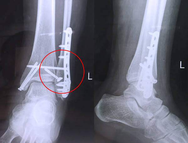
lateral malleolus fracture

posterior malleolus fracture
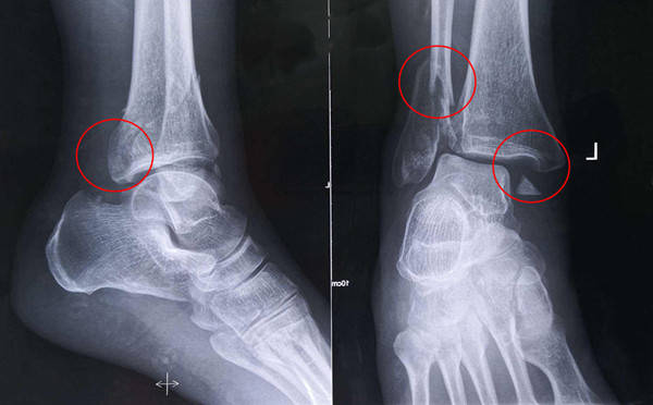
Three ankle fractures
【Medical Science】Pilon Fracture
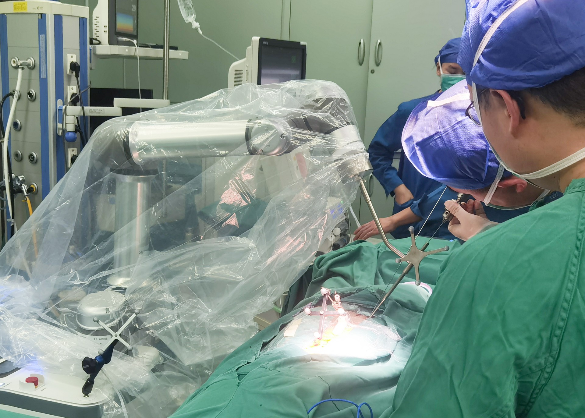
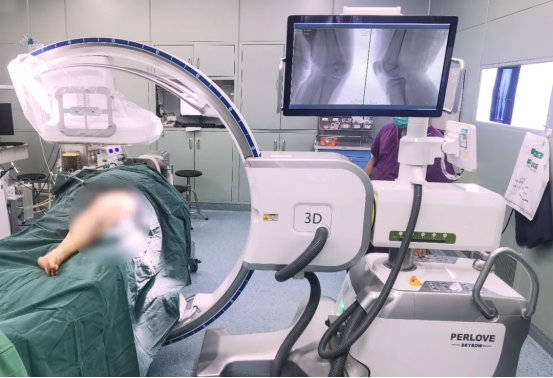
There is a special type of ankle fracture that may not show up on plain X-rays. It is when the articular surface of the tibia collapses inward. We also call it a "Pilon fracture". When it collapses in, we may not be able to see if it is collapsed on the plain X-ray, but we can see a 360° image of the ankle on CT, so we can see if the patient has a collapsed ankle fracture. In the same way, when we do Pilon fracture surgery, ordinary two-dimensional X images cannot judge the quality of our surgical reduction. At this time, if the doctor has a tool that can take three-dimensional images, then the doctor's accurate operation is Very beneficial.

*Individual differences, patients should be subject to hospital diagnosis.
3D Imaging Helps Accurate Treatment of Complex Surgery
-
Surgical Robots Take the Stage in the “Battle to Protect the Spine”
Read More » -
Application of 3D C-arm Imaging in Radiofrequency Ablation Treatment of Trigeminal Neuralgia
Read More » -
Correcting Limb Length Discrepancy | 3D C-arm Assisted Epiphysiodesis in Pediatric and Adolescent Patients
Read More » -
Perlove Medical Concludes a Successful Presence at RSNA 2025 in Chicago
Read More » -
Multiple C-arm Systems From Perlove Medical Installed at Zhujiang Hospital of Southern Medical University
Read More » -
Perlove Medical 3D C-arm Installed at Ningde City Hospital
Read More »


