2025-12-10 15:49:13 Views:925
When children and adolescents suffer from gait abnormalities, compensatory spine scoliosis and other issues due to lower limb length discrepancy, epiphysiodesis of the knee (figure-eight plate) emerges as a key option to improve their growth and development. With its professional 3D imaging, Perlove Medical’s 3D C-arm is applied in the entire surgical process. Centered on precise imaging, it overcomes the difficulties in surgical operations and provides reliable imaging support for bone correction in children and adolescents.
Screw placement is a critical step in the surgery. It requires accurate placement in specific epiphyseal regions of the femur and tibia, which not only achieves the growth inhibition effect but also avoids damaging healthy epiphyses, blood vessels, or nerves, imposing demanding exceptional surgical accuracy. When locating the target sites (such as the femur and tibia) , surgeons can clearly identify the position of the epiphyseal plate using Perlove Medical’s 3D C-arm. This helps them define the safe zones for drilling and screw insertion, establish an optimal relative relationship between the screws, epiphyses, and bone axes, and eliminate risks like screw deviation or bone penetration.

Preoperative Imaging
Intraoperative 3D images were acquired by the Perlove Medical’s 3D C-arm.


During the procedure, the positioning guidewire is inserted into the growth plate. A 3D scan is then performed by the 3D C-arm to confirm the guidewire's placement. Once verified, the plate is attached and secured with cannulated screws.

A final C-arm fluoroscopy check showed satisfactory positioning of the “eight-Plate” and proper screw direction. The wound was irrigated with saline, sutured, dressed, and immobilized with a brace, marking the completion of the procedure.


After screw placement, it is necessary to confirm whether their positions meet biomechanical requirements, as this directly affects the effectiveness of postoperative growth inhibition and the safety of skeletal development. Traditional 2D fluoroscopy cannot fully assess the screws’ positions in three-dimensional space, making it easy to overlook subtle deviations.
Perlove Medical’s 3D C-arm performs postoperative scans to verify screw placement from multiple dimensions: the relationship between the screws and the epiphyseal line
, any loosening or displacement and whether the femoral and tibial mechanical axes are approaching balance. If any subtle issues are detected, timely adjustments can be made to ensure optimal screw fixation. This postoperative 3D verification eliminates the observation blind spots in 2D imaging and serves as the final safeguard for surgical outcomes and long-term skeletal health.
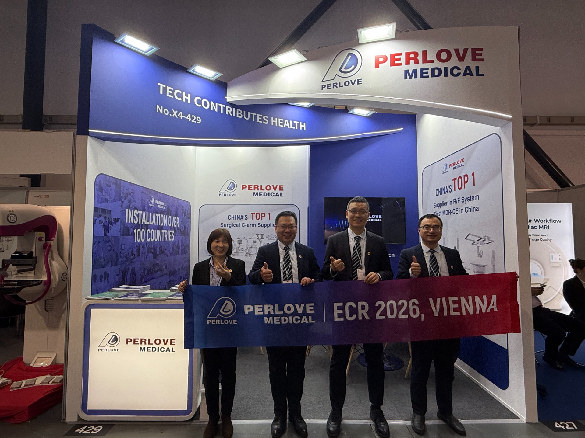
PERLOVE MEDICAL at ECR 2026 | Advancing Radiology Innovation in Vienna
Read More »Perlove Medical at ECR 2026: Showcasing Cutting-Edge Imaging Solutions in Vienna
Read More »🌍 Elevating Global Healthcare | WHX Dubai 2026 Successfully Concluded
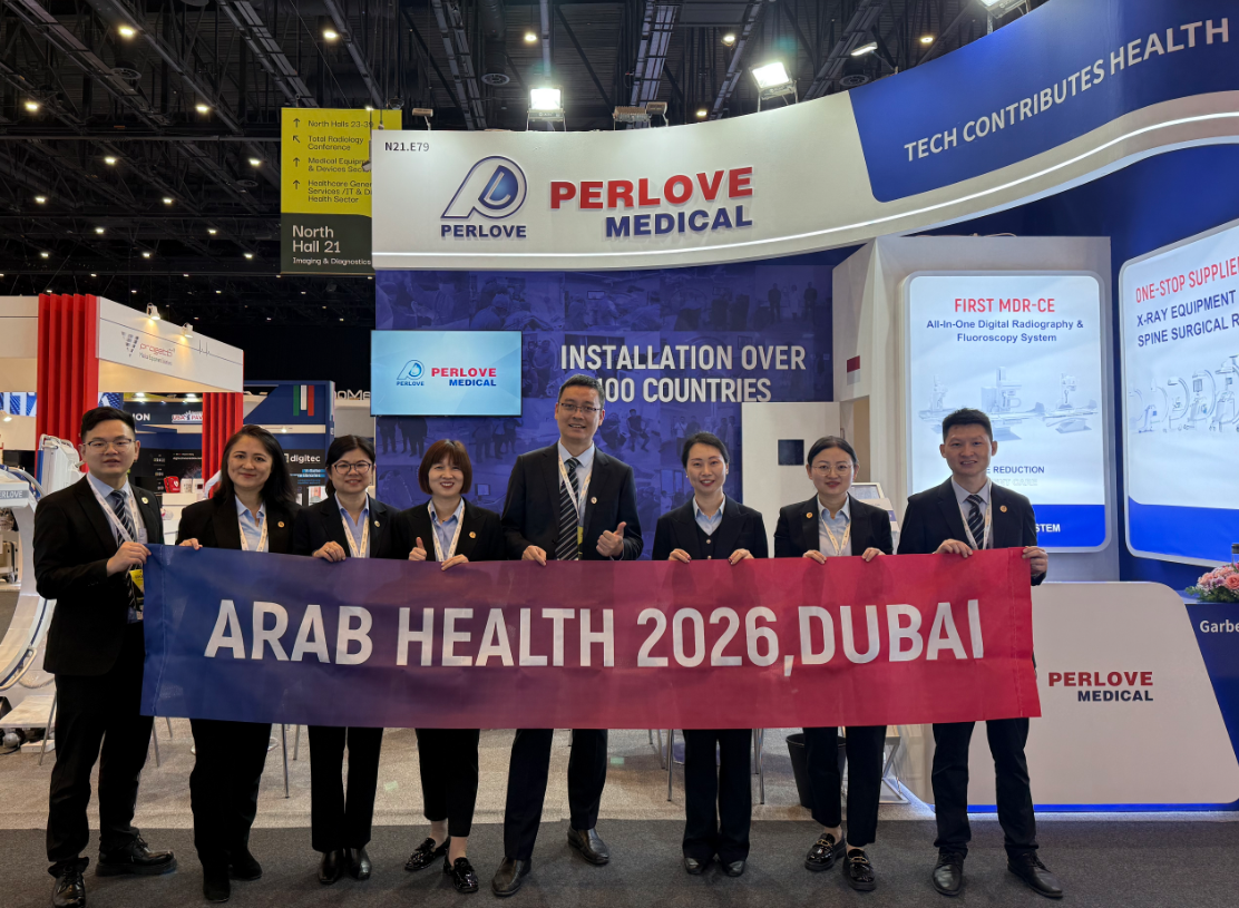
Read More »Join Us at ARAB HEALTH 2026 | See You in Dubai
Read More »Surgical Robots Take the Stage in the “Battle to Protect the Spine”
Read More »Application of 3D C-arm Imaging in Radiofrequency Ablation Treatment of Trigeminal Neuralgia
Read More »