News
2022-03-02 15:09:04 Views:1900
Case presentation: Clinical application of PLX7500 3D surgical C-Arm system
The mobile 3D surgical C-arm system which is a combination of conventional 2D radiography and CT-like imaging can creates 3D radiographic image consisting of transverse, sagittal and coronal planes. It brings a brand-new horizon of bone tissue and implants for surgeons, provides maximum guarantee for complex procedures as well as decrease of complications.
Case 1
Description: Female, 56 years old, suffers from lower back pain, combined with pain in lower limbs for 5 years, has limitation of movement. The pain deteriorates when patient engaged with prolonged standing or bending, and relieves when patient lies down. In the last one month, the situation has been worsened.
Diagnosis: MR image shows that L3/L4, L4/L5 disc herniation and intraspinal space-occupying lesion.
Treatment: Laminectomy at L5, S1 vertebrae + pedicle screw fixation
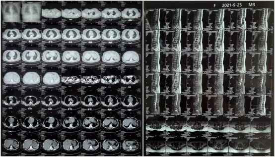
MRI shows disc herniation at L3/4/5 and intraspinal space-occupying lesion
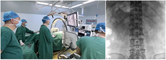
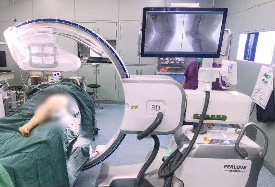
PLX7500 3D surgical C-arm system and presurgical radiography
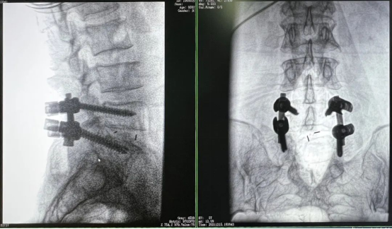
Pedicle screw implantation at L5 S1 guided by radiographic images
3D image achieved by C-arm rotatory scanning
3D radiographic image consisting of transverse, sagittal and coronal planes.
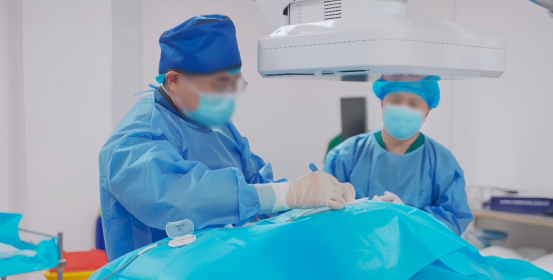
Suturing after surgery
Case 2:
Description: Male, 52 years old, confirmed with comminuted fracture at tibial plateau by presurgical 3D reconstruction.
Treatment: Open reduction and internal fixation
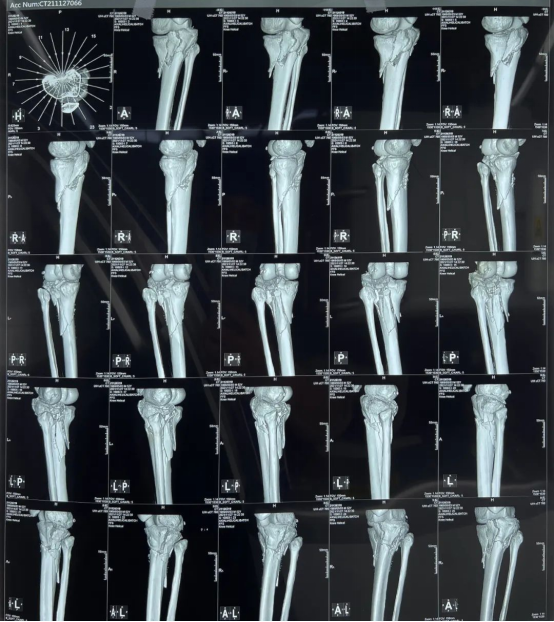
Comminuted fracture comfirmed by 3D reconstruction
Insert K wire and screws for fixation
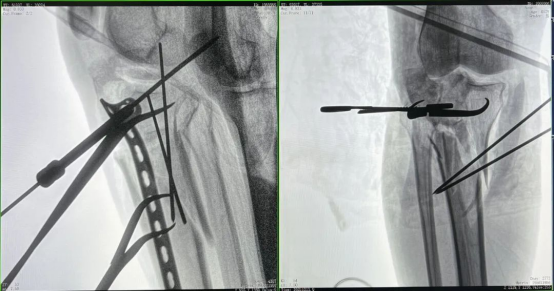
High definition image of AP and lateral view
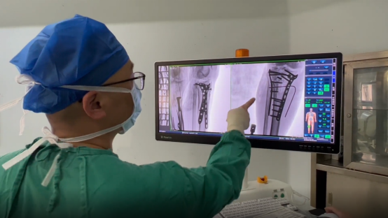
After-surgery confirmation
-
Surgical Robots Take the Stage in the “Battle to Protect the Spine”
Read More » -
Application of 3D C-arm Imaging in Radiofrequency Ablation Treatment of Trigeminal Neuralgia
Read More » -
Correcting Limb Length Discrepancy | 3D C-arm Assisted Epiphysiodesis in Pediatric and Adolescent Patients
Read More » -
Perlove Medical Concludes a Successful Presence at RSNA 2025 in Chicago
Read More » -
Multiple C-arm Systems From Perlove Medical Installed at Zhujiang Hospital of Southern Medical University
Read More » -
Perlove Medical 3D C-arm Installed at Ningde City Hospital
Read More »


