In orthopedic surgical treatments such as spine and long bone fractures, obtaining a larger field of view and clearer images of the fracture site can help doctors understand and assess the alignment and alignment of the fracture site in a timely manner intraoperatively, and enable length and angle measurements, providing strong support for further improving the quality of surgery.At the end of 2021, P.A.M. launched the PLX119C, an all-in-one C-arm with a wider field of view and clearer images for a better clinical experience!
Integrated large tablet C-arm orthopedic surgery case sharing
Patient: Male, age:43, Left femoral stem fracture
——Internal fixation of left femoral stem fracture with intramedullary nail
The femur is the main weight-bearing bone of the lower extremity and has its own special anatomical relationship with well-developed surrounding muscles. Therefore, once a femoral stem fracture is improperly treated, it is easy to cause deformity and dysfunction due to muscle strain. In the treatment of femoral stem fracture, it is important to restore the length and force line of the limb without rotation, and to protect the local blood flow of the fracture by minimally invasive means as much as possible to promote healing. Intramedullary nailing can achieve greater stability and sturdiness of the fracture, reduce medical contamination, reduce soft tissue separation and destruction of the surrounding blood supply, and facilitate early healing of the fracture, which is the preferred treatment for femoral stem fractures.
Pre-operative examination

C-arm assisted surgical procedure
Intraoperative fluoroscopy was performed using the large flat-panel Integrated C-arm machine of Perlove Medical to determine the fracture condition and the position of metal implants such as Kirschner wire and intramedullary nails, and to make adjustments.
The accurate intraoperative positioning of the C-arm greatly shortened the operation time, reduced the patient's pain, and assisted in the successful completion of the operation.


When performing intramedullary nail fixation, the surgeon needs to observe both the nail entry point and the fracture site. The large flat panel all-in-one C-arm of Perlove Medical adopts a 30CM×30CM flat panel detector, which can present a broader imaging area and meet most of the needs of long bone intramedullary nail fixation photography.

Intraoperative positioning was accurate, bleeding was low, and postoperative images showed correction of femoral displacement and restoration of good lower limb force lines.
Comparison of PLX119C clinical images with conventional images
Note: The blue dashed line shows the imaging area of the conventional 20CM x 20CM plate.


Perlove always committed to providing patients with better quality high-end medical equipment and more optimal clinical solutions to protect every equal and precious life!
Chat with us for more info!
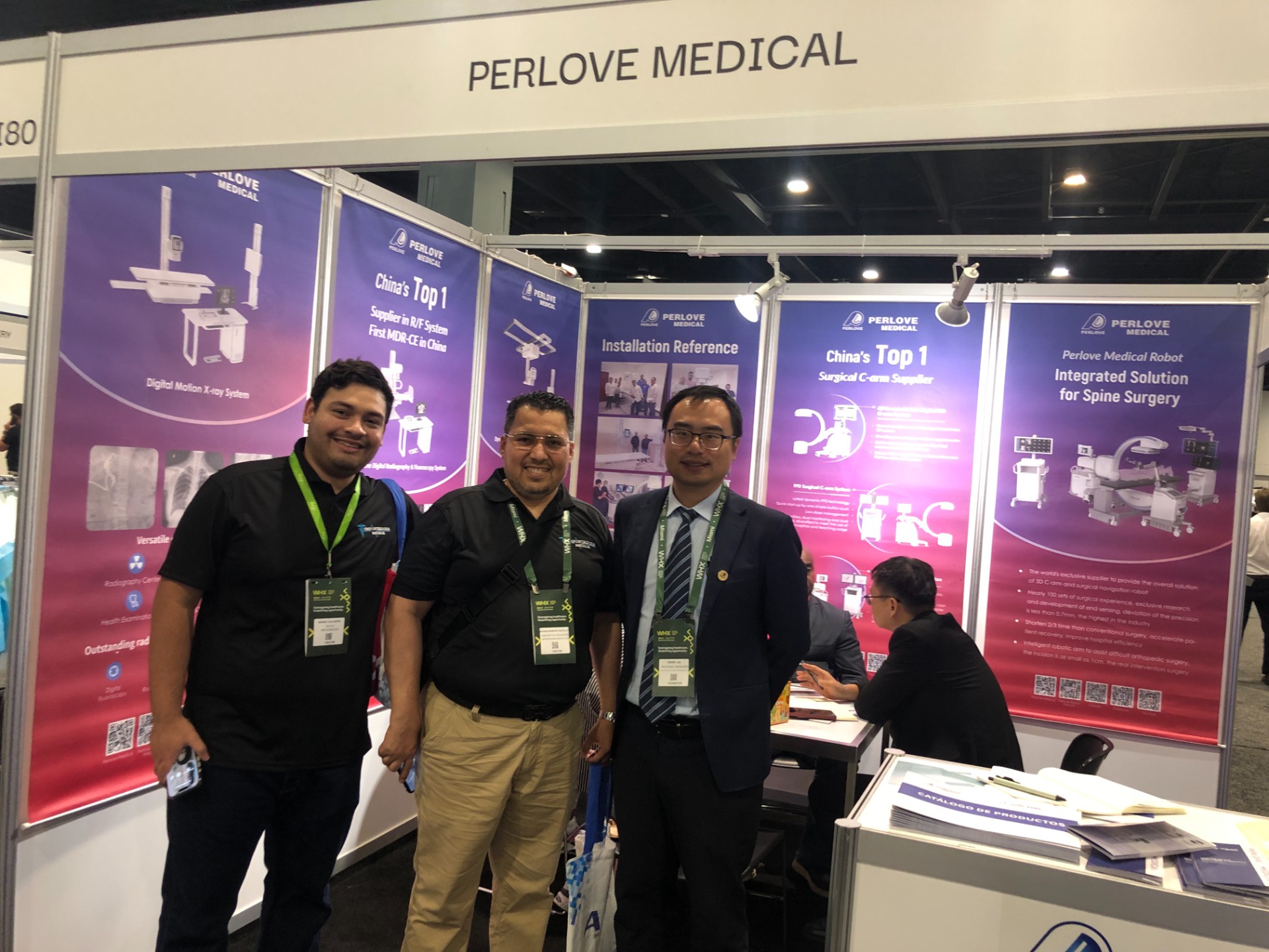
🌍 WHX Miami 2026 – Exhibition Wrap-Up
Read More »Perlove Medical to Exhibit at WHX Miami — See You in Miami!
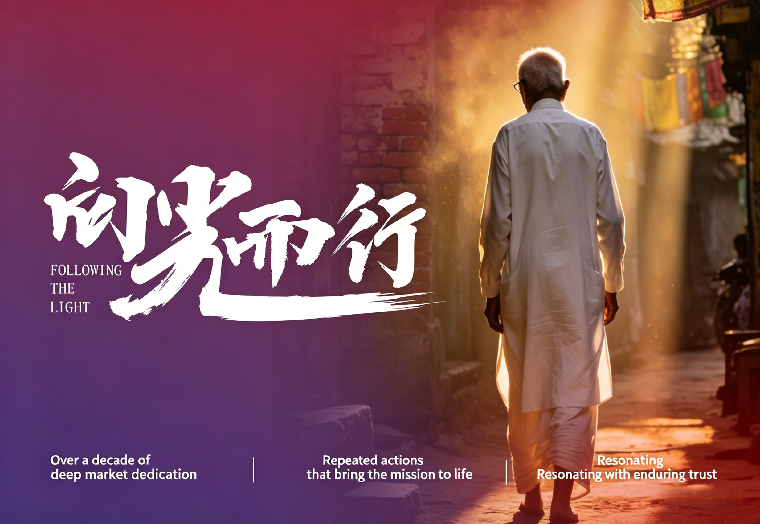
Read More »A Decade of Dedication, Echoing in the Market Again | Perlove Medical’s C-arms Deployed in Three Major Regional Hospitals in Bangladesh
Read More »2026 Innovate Together, Grow Further丨Perlove Medical International Distributor Promotion Conference
Read More »PERLOVE MEDICAL at ECR 2026 | Advancing Radiology Innovation in Vienna
Read More »Perlove Medical at ECR 2026: Showcasing Cutting-Edge Imaging Solutions in Vienna
Read More »