News
2022-08-09 14:39:36 Views:1831
Application of Perlove medical large plate integrated C-arm in urological double J-tube implantation
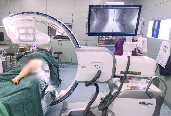
Recently, Director Wei Zhongqing of the Second Affiliated Hospital of Southern Medical University led his team to successfully perform a double J-tube placement procedure for a patient with ureteral stenosis. During the operation, the large-field C-arm of the Perlove Medical Group provided imaging guidance for the precise placement of the double J-tube and ensured a smooth operation.

Case name: Transurethral double J tube placement
Unit: The Second Affiliated Hospital of Nanjing Medical University
Patient's age: 85 years old
Patient's gender: female
The patient was an 85 year old grandmother who had previously undergone percutaneous nephrolithotomy (PCNL). After the operation, the doctor placed a double J tube (ureteral stent) to ensure that the patient could urinate normally due to ureteral stricture, which resulted in difficulty in urination and renal insufficiency.
What is ureteral stricture?
The ureters are a pair of long, thin lumenal channels that connect the kidneys to the bladder. Their main function is to drain urine from the kidneys into the bladder. When the ureters become narrowed, it can lead to dilated ureters and hydronephrosis, which can lead to symptoms such as pain, urinary tract infections, haematuria and kidney stones. Factors that may trigger ureteral strictures include kidney stones, congenital disorders, medically induced injuries, radiation therapy, and malignancy. Studies have shown that although there is only a 3.5% chance of developing ureteral stenosis after lumpectomy, nearly 75% of ureteral stenosis is caused by medical injury and radiation therapy. Patients are 85 years old and have a relatively high probability of post-operative complications.

The role of the double J tube
There are a number of treatment strategies for ureteral strictures, including open surgery, trans-laparoscopic ureterectomy and nephrostomy. The main goal is to create a new channel for the urine to pass out of the kidney. The double-J tube, also known as the D-J tube, is a common ureteral stent with pigtailed curved ends that is placed in the ureter to both drain urine and prevent it from slipping into the kidney or bladder. Double J-tube placement, a minimally invasive treatment modality, is one of the most common options for achieving ureteral evacuation, with a shorter procedure time and less trauma to the patient than other treatment strategies.

The procedure
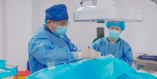
The patient underwent this procedure using the stone position. After general anaesthesia, the ureteroscope is operated to enter the bladder through the urethra, and the ureteral opening is located by microscopic observation. At this point, a guidewire is inserted under fluoroscopic guidance of the large flat C-arm of the Perlove Medical, which reaches the renal pelvis and confirms the position, while the ureteroscope is used to locate the stricture. Next, the double J-tube stent is inserted along the guidewire, again under fluoroscopic guidance, and when the two ends of the stent reach the appropriate position, the guidewire and ureteroscope are withdrawn to complete the procedure.


Summary of surgical difficulties and technical advantages
Large field of view: more detail at once

When using a conventional shadow-augmented C-arm or a 9-inch flat detector, the imaging area is small, and when positioning the guidewire for observation, it is not possible to present a large fluoroscopic image due to the limitations of the surgical bed, which requires several shots, thus reducing the efficiency of the procedure. The large flat panel C-arm is equipped with a 12" flat panel detector, which compensates for the difficulty in positioning.
-
Surgical Robots Take the Stage in the “Battle to Protect the Spine”
Read More » -
Application of 3D C-arm Imaging in Radiofrequency Ablation Treatment of Trigeminal Neuralgia
Read More » -
Correcting Limb Length Discrepancy | 3D C-arm Assisted Epiphysiodesis in Pediatric and Adolescent Patients
Read More » -
Perlove Medical Concludes a Successful Presence at RSNA 2025 in Chicago
Read More » -
Multiple C-arm Systems From Perlove Medical Installed at Zhujiang Hospital of Southern Medical University
Read More » -
Perlove Medical 3D C-arm Installed at Ningde City Hospital
Read More »


