News
2022-08-24 14:55:24 Views:1072
Lumbar posterior interbody fusion technology training camp: Perlove integrated mobile C-shaped arm to help the operation
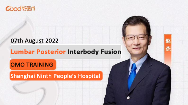
On August 7, the Good Medicine Training Camp invited orthopedic professionals - Shanghai Jiaotong University Medical School Affiliated Ninth People's Hospital Professor Zhao Jie for in-depth explanation and guidance of lumbar posterior interbody fusion technology, through the standardized (nail placement, decompression range, interbody fusion preparation) operation process, refine the technology, improve the treatment effect The course will help doctors learn the knowledge and surgical skills of lumbar posterior interbody fusion technology systematically and efficiently in a short period of time.
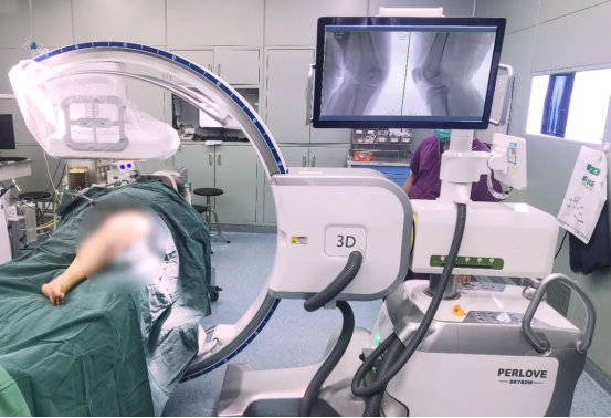
Perlove Medical large plate integrated mobile C-shaped arm to help the operation
Lumbar Interbody Fusion (LIF) is a traditional procedure to treat lumbar instability by joining or fusing two or more vertebrae together. It is mainly used in cases of destructive vertebral disease, spinal instability, prevention or correction of deformity, etc. It has been successfully applied to relieve low back pain, rebuild spinal stability and restore the normal sequence of the spine, and is by far one of the most important surgical procedures in spine surgery to treat serious diseases of the lumbar spine.
Posterior lumbar interbody fusion (PLIF) is one of these surgical procedures. It is performed through an incision in the back of the body to expose the spine. The disc is removed, an intervertebral fusion is used to maintain proper disc height, and a bone graft is used to promote fusion of adjacent vertebrae and maintain spinal stability.
Surgical steps
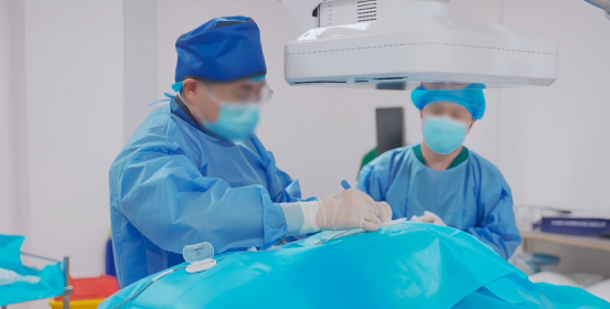
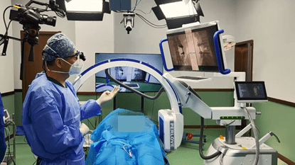
The incision and field are revealed by making an incision of approximately 8-10 cm at the back of the back. The spinal site to be operated on is accurately positioned using a large flat mobile C-arm from Perlove
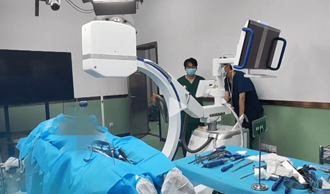
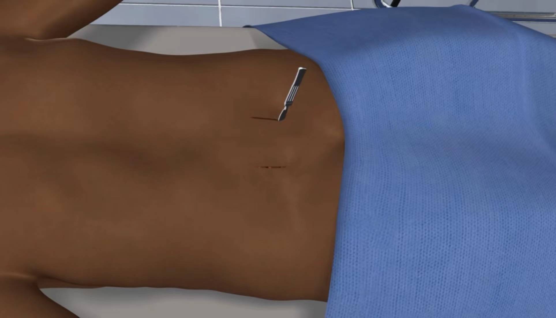
The skin and subcutaneous tissue are incised, and the paravertebral muscles are peeled away to fully expose the upper and lower vertebral segments, and laterally to fully expose the transverse processes, which are propped open with a deep automatic spreader and compressed to stop bleeding and create space for surgery.
The arch plate (vertebral plate) is carefully removed so that the nerve root can be seen and accessed. The small joint above the nerve root may be trimmed to allow more space for the nerve root. The surgeon then removes the affected disc and its surrounding tissue and prepares the surface of the adjacent vertebrae for fusion.
Between the adjacent vertebrae, a structural support cage (interbody fusion) made of titanium, carbon fiber interbody fusion or polymer is implanted to maintain the vertebral space height. The interbody fusion removes pressure from the compressed nerve roots and preserves the proper intervertebral space height to provide stability to the spine and support normal loading. The intervertebral fusion is often filled with autogenous bone, or allogeneic bone...to promote bone regeneration and permanently fuse the vertebrae together.
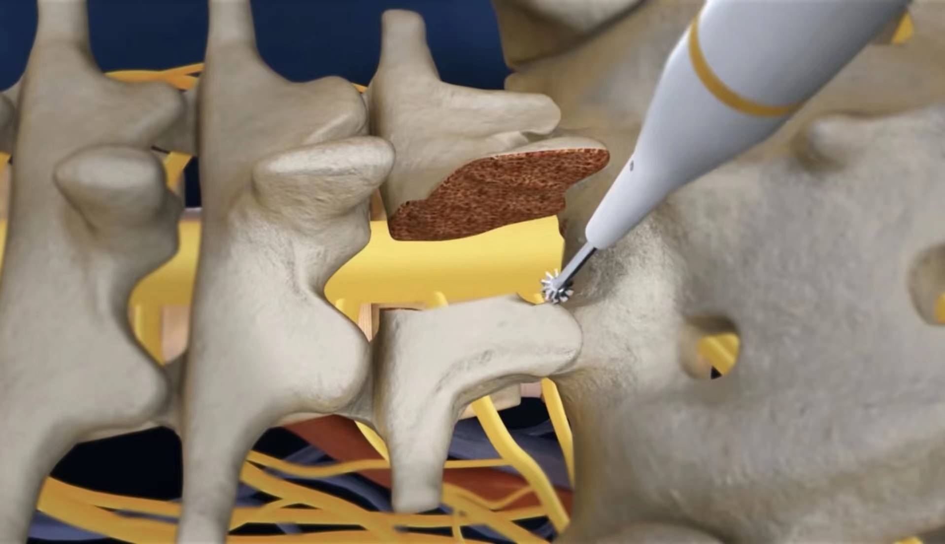
Arch nail screw insertion: Bite off the cortex at the entry point with a bone biting forceps, prepare the screw entry point and orifice with an opener, and insert the positioning pin.
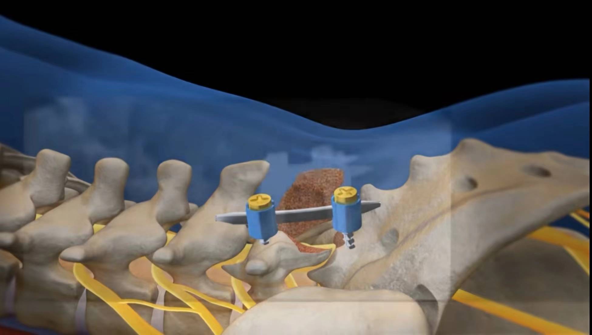
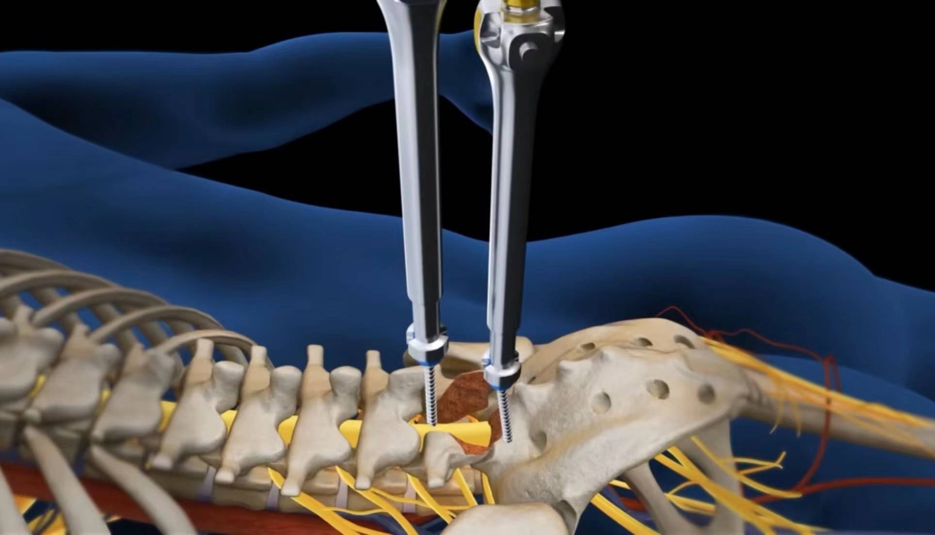
The standard screw is implanted with a one-piece mobile C-arm fluoroscopy to confirm correctness. Insert the remaining screws in the same way. Confirm the screw position and length using C-arm fluoroscopy again. Use the rod holder to insert the desired length and bend of the nail rod. The nut is first temporarily locked, and the bracing forceps and bar holder are used to create bracing and compression between the collets before the nut is finally tightened to avoid any loosening and wobbling.
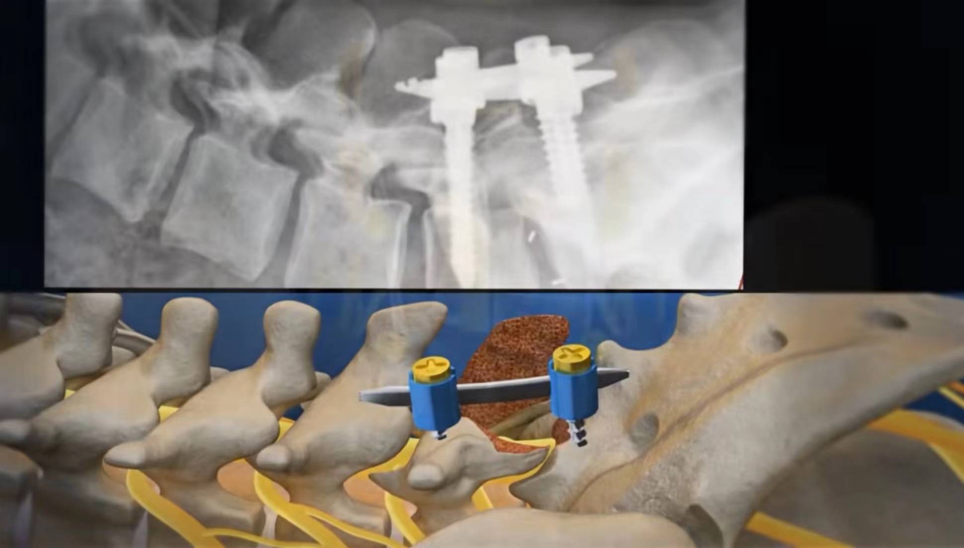
The instruments are removed, the incision is closed and bandaged to complete the procedure.
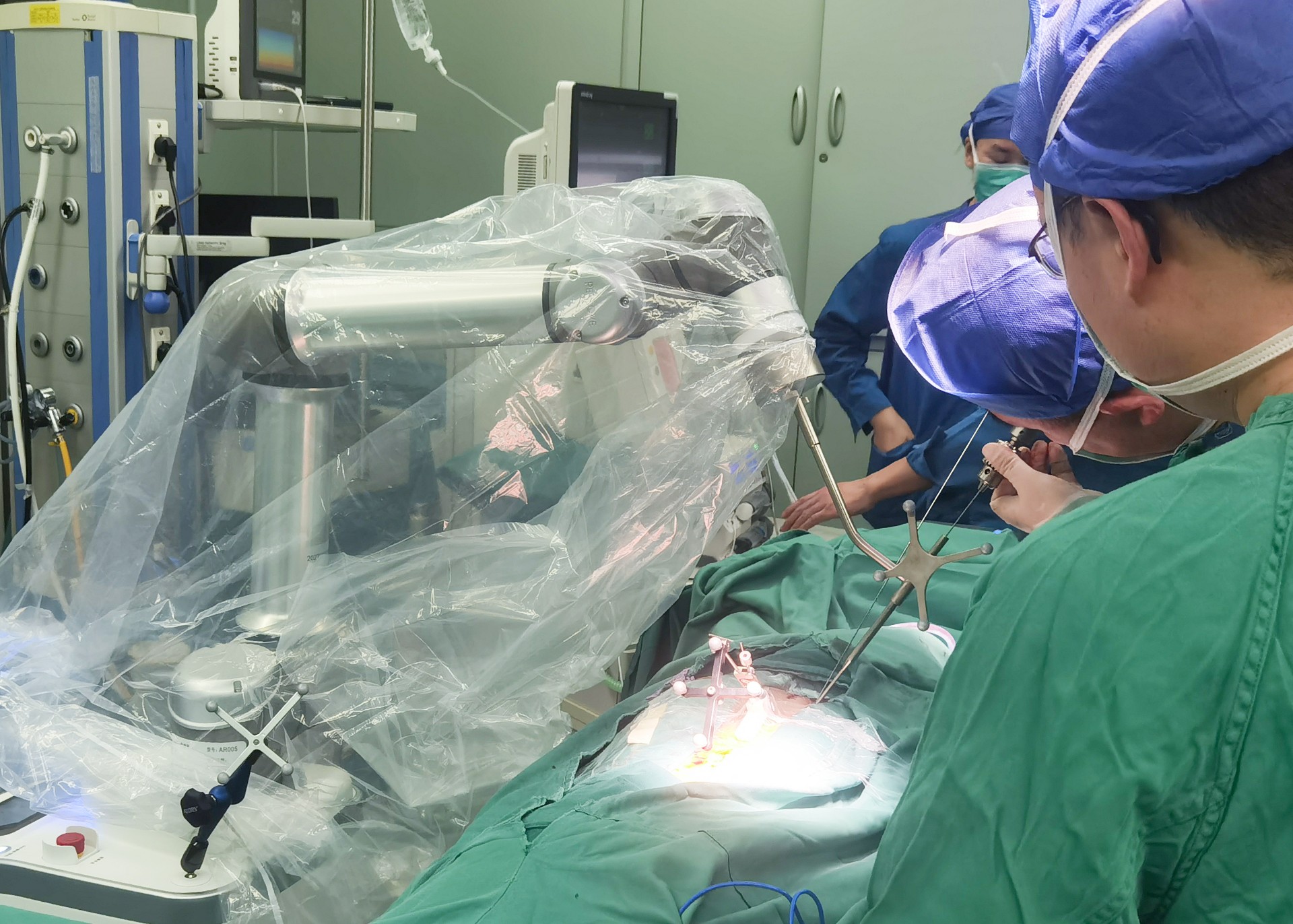
The Perlove Medical large flat panel all-in-one mobile C-arm uses a 30CM x 30CM flat panel detector, which can generally image 5 lumbar vertebrae at one time, presenting a broader field of view. It enables the surgeon to observe the vertebral fluoroscopy comprehensively at one time, making the surgery more efficient and accurate.
If you want to know more about the introductio, welcome to chat with us!
-
Surgical Robots Take the Stage in the “Battle to Protect the Spine”
Read More » -
Application of 3D C-arm Imaging in Radiofrequency Ablation Treatment of Trigeminal Neuralgia
Read More » -
Correcting Limb Length Discrepancy | 3D C-arm Assisted Epiphysiodesis in Pediatric and Adolescent Patients
Read More » -
Perlove Medical Concludes a Successful Presence at RSNA 2025 in Chicago
Read More » -
Multiple C-arm Systems From Perlove Medical Installed at Zhujiang Hospital of Southern Medical University
Read More » -
Perlove Medical 3D C-arm Installed at Ningde City Hospital
Read More »


