News
2022-09-13 14:10:59 Views:1618
What are the advantages of the large size FPD C-arm? Display of large FPD images
At present, with the further development of flat panel detector related technologies, the C-arm X-ray machine imaging system is gradually upgraded from an image intensifier to a digital X-ray flat panel detector. The effective imaging area of the flat panel C-arm is larger and the imaging quality is higher, which can better meet the needs of clinical use and become the new favorite of the C-arm products of radiographic imaging equipment.
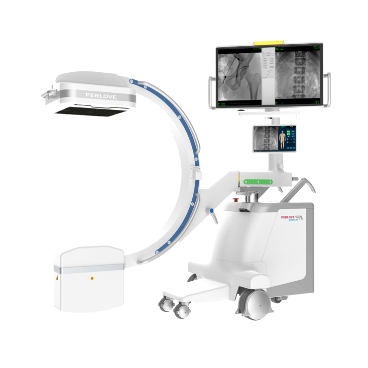
Perlove PLX119c is a large size FPD C-arm, which brings a wider field of view and clearer images to the clinic.
So, what are the advantages of the large size FPD C-arm?
Advantages of FPD:
1. By virtue of the advantages of digital imaging, the flat panel detector avoids the defects such as signal conversion loss, geometric distortion, color interference and high inconsistency in the imaging of the image intensifier. At the same time, the full digital image is convenient for transmission, storage and printing, and its service life is longer than that of the image intensifier.
2. Compared with the 10bit / 12bit gray-scale imaging provided by ordinary image intensifier, the flat-panel C-arm X-ray machine can provide up to 16bit gray-scale imaging, which makes the image more hierarchical and can see more details to facilitate the clinical use.
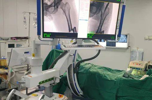
Advantages of large-size FPD:
The large plate C-arm is configured with 30 * 30cm amorphous silicon FPD, providing a wider field of view. Compared with the traditional 9-inch image intensifier, the imaging range is increased by 1.6 times. One exposure can image the whole lumbar spine, covering the entire pelvic range. It is convenient for doctors to better locate the focus and plan the operation , which can reduce the inconvenience caused by multiple exposures due to insufficient FOV and improve the operation efficiency.
Comparison of PLX119c clinical image and traditional image
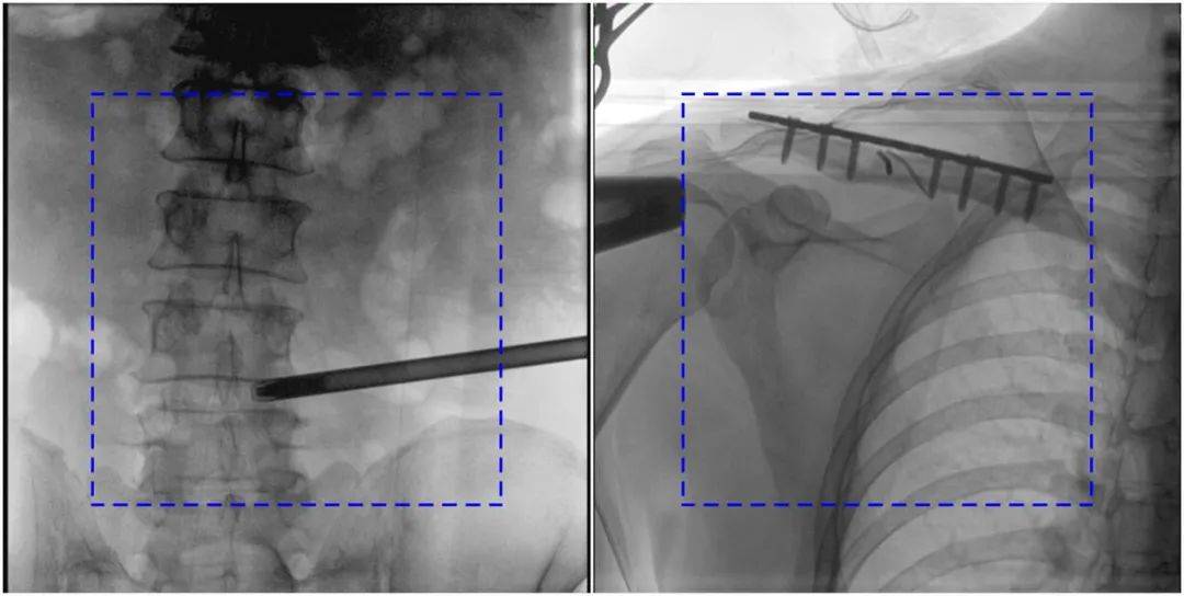
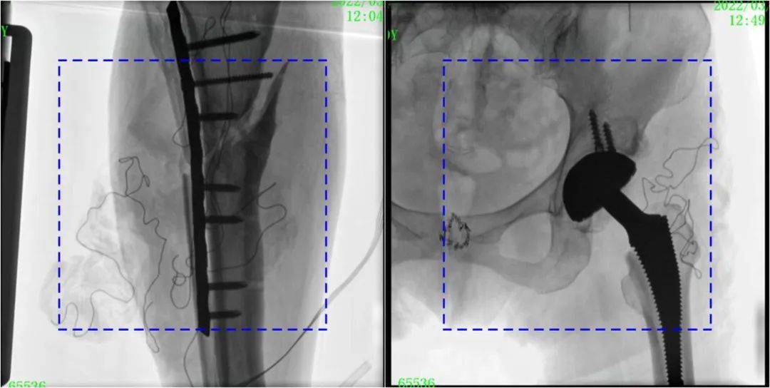
The blue dotted line is the traditional 21cm ×21cm image
Most of the main C-arm system in the market with 21cm × 21cm FPD or image intensifier has a small imaging range and can generally image 3.5 lumbar vertebrae. It may be necessary to take multiple shots to determine the injured vertebrae. Perlove Medical large FPD C-arm adopts 30cm×30cm flat panel detector can generally image 5 lumbar vertebrae at one time, presenting a broader field of vision. So that the doctor can observe the injured vertebral body and the surrounding vertebral body comprehensively with one exposure, making the operation more efficient and accurate.
-
Surgical Robots Take the Stage in the “Battle to Protect the Spine”
Read More » -
Application of 3D C-arm Imaging in Radiofrequency Ablation Treatment of Trigeminal Neuralgia
Read More » -
Correcting Limb Length Discrepancy | 3D C-arm Assisted Epiphysiodesis in Pediatric and Adolescent Patients
Read More » -
Perlove Medical Concludes a Successful Presence at RSNA 2025 in Chicago
Read More » -
Multiple C-arm Systems From Perlove Medical Installed at Zhujiang Hospital of Southern Medical University
Read More » -
Perlove Medical 3D C-arm Installed at Ningde City Hospital
Read More »


