Esophageal cancer, also called esophageal cancer, is a malignant tumor occurring in the epithelial tissue of the esophagus, accounting for 2% of all malignant tumors, and approximately 220,000 people die from esophageal cancer worldwide each year. China is the country with high incidence of esophageal cancer in the world and one of the countries with high mortality rate of esophageal cancer in the world. Esophagogram is the basic examination method for esophageal lesions, which can detect the characteristic changes of esophageal cancer - interruption and destruction of esophageal mucosa, and patients often feel dysphagia, and this feature is the most common in clinical practice and typical manifestation of early esophageal cancer. The general accompanying features include filling defect of the wall, niche shadow, soft tissue mass shadow, narrowing of esophageal lumen, etc.
Why is dynamic DR used in esophagography?

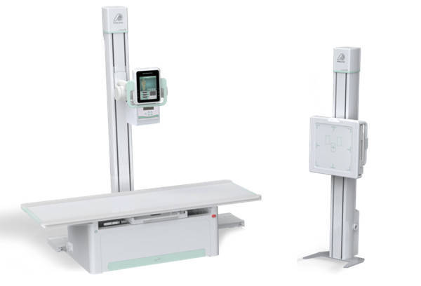
Dynamic DR
Dynamic DR rectangular acquisition area is large, no need to move the bulb and detector up and down, one exposure can show the whole esophagus; fast, real-time HD spot film technology can take in key image information and clearly show the lesion characteristics, which provides great support for diagnosis.
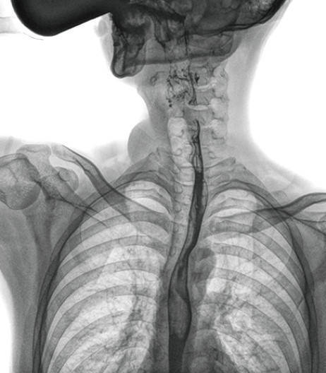
Compared with the past digital gastrointestinal machine, dynamic DR image resolution is high. In the panoramic view of the esophagus, local mucosal destruction, interruption, luminal narrowing and the extent of the lesion are displayed with significantly better clarity than the gastrointestinal machine.
Esophageal cancer is often not recommended for proposed esophagoscopy because of luminal narrowing, and the use of fiberoptic esophagoscopy often has the probability of increasing patient discomfort, inconvenient passage of the tube and even mucosal bleeding. After applying dynamic DR, the proposed esophagogram can dynamically observe the clinical signs of the canal wall, which is easier to clearly show the lesion characteristics of esophageal cancer and clarify the lesion scope, and has certain guidance significance for surgery. In the presence of dysphagia, two clinical approaches are often applied to distinguish benign esophageal tumors, cardia achalasia and benign esophageal strictures from esophageal cancer.
Pratt & Whitney Dynamic DR can show the whole esophagus in one exposure
Dynamic DR is capable of large-format fluoroscopy, instantaneous high-resolution spot film, etc. In esophagography, it is convenient to observe esophageal lesions in one large format because of the very fast flow rate of contrast after swallowing barium. With instantaneous spot film, the image of the lesion can be captured in real time so that a quick diagnosis can be made. With a large field of view of 17×17 inches, Pride Dynamic DR can display the entire esophagus in one exposure, which is more convenient to observe the lesions of the esophagus and determine the extent of the lesions, which is an important reference value for diagnosis and treatment.
Dynamic DR can dynamically observe whether the peristalsis of the canal wall is stiff to identify benign and malignant strictures, not only in the fluoroscopy process, real-time high-definition spot film, to achieve millisecond dynamic and static image switching, quickly capture the image of the lesion site, imaging clear and rapid, as far as possible to reduce the time of patients with functional esophageal disorders due to swallowing difficulties and endure pain, while improving the efficiency of doctors to make the correct diagnosis, but also real-time preservation of Video images can be repeatedly observed and analyzed to clarify the extent of the lesion, which is an important guide for surgery.

Dynamic DR in the upper gastrointestinal imaging image
It is easy to see that the clinical application of dynamic DR in esophagography, compared with other means of examination, clear imaging, easy to apply, and can fully display the local and overall structure of the esophagus morphology, and reveal the relevant morphological and functional changes, more conducive to help achieve accurate diagnosis.
It can be used in various clinical departments, such as physical examination, internal medicine, surgery, orthopedics, emergency medicine, etc. It is a multi-functional machine that can be used for routine filming, gastrointestinal, fluoroscopy and imaging, which can meet the radiological needs of primary hospitals.
-
Another International Milestone! Multiple Perlove Medical Devices Achieve MDR CE Certification
Read More » -
Discover Innovation at Medic East Africa 2025 in Nairobi, Kenya!
Read More » -
JFR 2025 in Paris Concludes Successfully – Perlove Medical Highlights
Read More » -
Orthopedic Robotic-assisted MIS-TLIF surgery
Read More » -
Discover Innovation at JFR 2025 in Paris, France!
Read More » -
【Respiratory Interventions, 3D Imaging Guidance】3D C-Arm Assists in Minimally Invasive Diagnosis and Treatment of Pulmonary Nodules Under Robotic Navigation
Read More »


