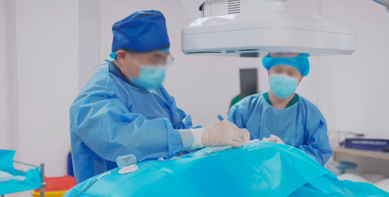
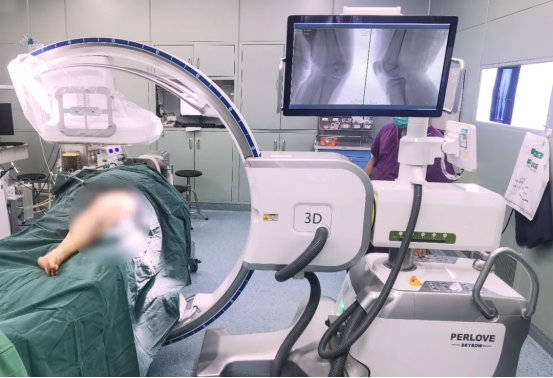
With the development of surgical techniques and imaging, the application of minimally invasive techniques in pelvic fractures is becoming more and more widespread. Minimally invasive surgical treatment of pelvic fracture is a screw fixation technique with a small incision, i.e. limited minimally invasive surgery, after closed repositioning under 3D flat-panel C-arm fluoroscopy. This method has the advantages of less surgical trauma, less patient pain, shorter operation time, faster patient recovery after surgery, and fewer surgical complications. It can greatly reduce the patient's pain.
Pelvic fracture is a serious injury, mostly seen in traffic accidents or injuries from falls, industrial accidents, etc. Since the body weight is transmitted through the pelvis to both lower limbs, and there are several organs and rich blood vessels and nerves in the pelvic cavity, pelvic fracture usually accompanies the injury of pelvic cavity view, blood vessels and nerves, in addition to directly causing functional impairment.
The treatment of pelvic fracture has always been a difficult task in traumatic orthopedics. The anatomical structure of the pelvis and its adjacent relationships are complex, and the pelvis has rich vascular and complex neurological anatomical features, and the anatomical form of the pelvis is irregular, so pelvic fracture surgery is difficult and one of the complex and challenging operations in the field of orthopedics.
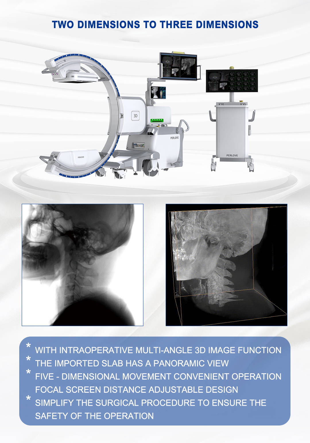
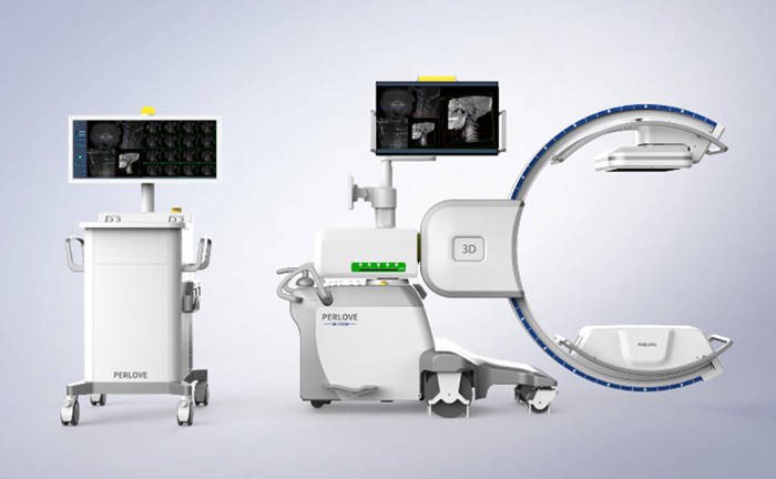
The Perlove Medical 3D Flat Panel C-Arm, with 190° of comprehensive information acquisition, acquires 100 or 380 frames of images by means of pulsed fluoroscopy, and volumetrically reconstructs them into 3D stereoscopic images, as well as the corresponding sagittal, coronal, and cross-sectional views.
Application of 3D flat-panel C-arm imaging in pelvic fracture surgery.
3D flat-panel C-arm images provide CT-like slice images (sagittal, coronal, and cross-sectional images), and multiple fluoroscopic position images can be viewed at the end of a single 3D scan without the need for multiple scans.
The 3D flatbed C-arm image contains richer information on the external shape of the bone and other anatomical structures, allowing visual evaluation of whether the screw is in the sacral nerve root foramen, the position of the screw in relation to the vertebral body (information on the LC-2 screw channel, the full length of the LC-2 screw, and the LCD line are also visually visible) and visual determination of the effect of pelvic compression fracture repositioning.
The large field of view, Perlove Medical 3D flat panel C-arm uses a 30cm x 30cm flat panel detector, which can present more image details and has application advantages for procedures such as bilateral pelvic fracture types or internal fixation of the posterior pelvic ring, where images of all fracture sites can be obtained with a single exposure.
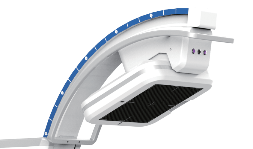
The key to a successful minimally invasive surgery is not only the surgeon's excellent medical skills, but also high-tech equipment to help. The 3D flat panel C-arm can show the anatomical structure of the pelvis and the relationship between fractures in a more realistic way, forming clear and accurate 3D images, which are of high value for determining the type of pelvic fracture and deciding the treatment plan.
-
Application of 3D C-arm Imaging in Radiofrequency Ablation Treatment of Trigeminal Neuralgia
Read More » -
Correcting Limb Length Discrepancy | 3D C-arm Assisted Epiphysiodesis in Pediatric and Adolescent Patients
Read More » -
Perlove Medical Concludes a Successful Presence at RSNA 2025 in Chicago
Read More » -
Multiple C-arm Systems From Perlove Medical Installed at Zhujiang Hospital of Southern Medical University
Read More » -
Perlove Medical 3D C-arm Installed at Ningde City Hospital
Read More » -
Perlove Medical Debuts New Products at the 2025 Chinese Conference of Digestive Endoscopology
Read More »


