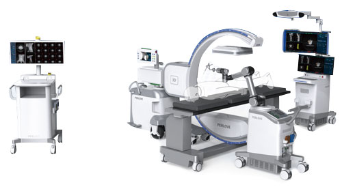
PLX C7600 Series
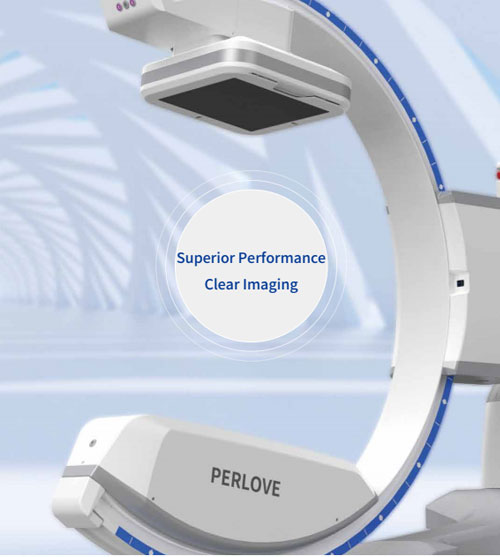
Digital Intraoperative 3D C-arm System
Digital Intraoperative 3D C-arm System Digital Intraoperative 3D C-arm System

3D Imaging, More Comprehensive | Intraoperative CT-like examination, Lower revision risk In joint surgery, implants misalignment is not easy to detect intraoperatively and the damage to cartilage is irreversible. If it is detected in postoperative CT, revision surgery is inevitable, increasing the probability of complications and infection risk. With intraoperative 3D imaging, implant misalignment can be detected immediately, reducing the need for a second surgery.
PLX C7600 Series 3D Mode – Cervical vertebrae |
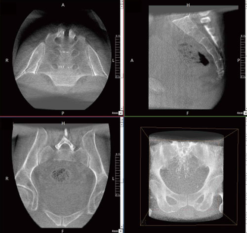
3D lmagingMore DetailsLarge 3D field of view (FOV) The 3D imaging FOV can cover BROAD VISIONLumbar vertebrae One 3D image can cover several vertebrae.
| STRUCTURE FULLY DISPLAYEDSacroiliac joints The bilateral sacroiliac and the entire internal fixation of the sacroiliac screws can be fully displayed in one 3D image.
|
Easy Positioning Fast Scanning
| ● Large free space, isocentric design, quicker positioning and collision checking Fast Scanning ● Multiple 3D acquisition modes, flexible selection High-speed Imaging ● Only 8 seconds, 3D reconstruction |
Intraoperative “CT” Imaging | The conventional process requires preoperative localization in the CT room before transfer to the operating room for thoracoscopic surgery, with the risk of pneumothorax, pulmonary hemorrhage, and guide needle displacement during transport. With the mobile 3D C-arm, 3D images of the lung can be acquired in the operating room. Under stable breathing, lung nodules can be precisely localized at sub-millimeter level, and the important anatomical structures (blood vessels, trachea, etc.) can be avoided effectively so that the intraoperative puncture can be safe and effective. Additionally, the mobile C-arm can significantly reduce the radiation dose and minimize X-ray damage to doctors and patients.
|
Advanced Imaging | The advanced dynamic liquid cooling system enhances the thermal capacity and heat dissipation efficiency of the tube, significantly extending the continuous working time of the tube to meet the highly frequent use.
|
| The field of view (FOV) is increased by 100% compared with the 9-inch flat panel, which helps the surgeon to quickly localize the vertebrae and the surgical site, avoiding multiple positioning and repeated exposures due to insufficient FOV. Ultra-wide Monitor The 32-inch monitor has a real-time/reference dual screen, making it easy to read the high-resolution film. 4-Megapixel Imaging Equipped with 4-megapixel flat panel detector, the 3D C-arm makes clinical images clearer and easy to observe tiny lesions, ensuring high-quality imaging of complex bonelacuna and soft tissues, such as chest, abdomen, spinal joints and so on. Intelligent Image Post-processing The workstation system processes the captured images and can intelligently recognize human tissues and metal implants. In addition, the system processes the image through artifact processing, tissue equalization, recursive noise reduction, etc., so as to present the images of surgical regions of interest (ROI) clearly. |
User-friendly Interaction |
The FPD can be flexibly adjusted, raised to increase the opening distance of the C-arm and lowered to get closer to the exposed area to expand the FOV.
Large Free Space The maximum space of 94 cm meets the positioning needs of obese patients or thicker tissues and reserves sufficient operation space for the surgeon.
|
LESS RADIATION, MORE CARERemovable Grid For dose-sensitive people (pediatric, etc), the filter grid can be easily and manually pulled out, which can save time and reduce the exposure dose.
Virtual CollimatorIt is able to adjust and preview the FOV in a ray-free state to reduce radiation damage from unnecessary exposure testings.
DAPThe intelligent DAP monitoring system can record the single examination dose and manage the radiation absorption of patients.
Low Dose ModeLow dose mode can significantly reduce the dose intake and unnecessary radiation, suitable for a variety of clinical examination needs. |
Additional FiltrationFour-mode additional metal filters are available to effectively filter soft X-rays from the initial radiation, improving imaging clarity while reducing patient radiation absorption.
Intelligent Dose SettingThe model has automatic adjustment of dose parameters according to the body parts. It can reduce radiation absorption while ensuring high-quality images.
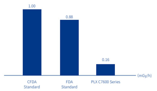
Lower leakage radiation in the loading stateLeakage radiation in the loading state of X-ray tube is far more lower than CFDA and FDA standard, providing better protection from the X-ray source.
Intelligent Pulse FluoroscopyPulsed X-rays are radiated at high power to obtain high-contrast images at the minimized radiation dose. |
Seamless Transmission with Navigation or Robotic SystemsIntegrated with a standardized data transmission interface for navigation or robotic systems, Navigation or robotic systems require the input of intraoperative 3D images, which can be seamlessly transmitted to the navigation system after image acquisition, containing high-resolution | The combination of 3D images and navigation and positioning systems can accurately complete implant positioning, especially in complex minimally invasive surgery, which can effectively reduce surgical incisions and bleeding, reduce the need for revision surgery and postoperative CT scans, and improve surgical efficiency. INTELLIGENT IMAGING
|
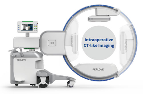
Comprehensive observation, Precise assessment, Safer surgery
The 3D C-arm can generate intraoperative transverse, sagittal, coronal and 3D images on real time to assess the relative positions of implants and anatomical structures in any slice from any angle.
The surgical results can be pre-confirmed in the operating room, which can be applied in all parts of the body.
Intraoperative CT-like examination, Lower revision risk
In joint surgery, implants misalignment is not easy to detect intraoperatively and the damage to cartilage is irreversible. If it is detected in postoperative CT, revision surgery is inevitable, increasing the probability of complications and infection risk. With intraoperative 3D imaging, implant misalignment can be detected immediately, reducing the need for a second surgery.
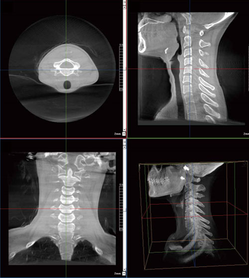
PLX C7600 Series 3D Mode – Cervical vertebrae
The 3D imaging FOV can cover
● All lumbar vertebrae
● All cervical vertebrae
● 8 thoracic vertebrae
● Most of the pelvis
● Unilateral acetabular joints
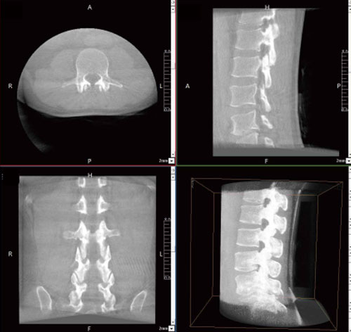
Lumbar vertebrae
One 3D image can cover several vertebrae.
The bilateral sacroiliac and the entire internal fixation of the sacroiliac screws can be fully displayed in one 3D image.
Easy Positioning
● Large free space, isocentric design, quicker positioning and collision checking
●Three-way positioning laser light, easier positioning of the patient site
●Position memory, increase first-time-right positioning for more efficient workflow
Fast Scanning
● Multiple 3D acquisition modes, flexible selection
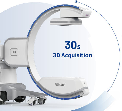
● 30-second fast acquisition, improved efficiency
High-speed Imaging
● Only 8 seconds, 3D reconstruction
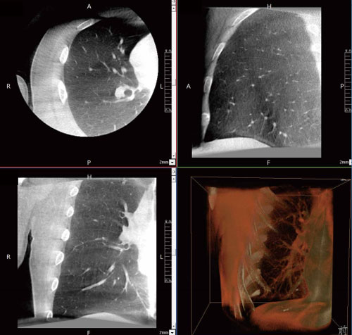
Meet the intraoperative imaging requirements for various procedures such as lung nodule biopsy, lung nodule resection, and lung nodule ablation treatment.
The conventional process requires preoperative localization in the CT room before transfer to the operating room for thoracoscopic surgery, with the risk of pneumothorax, pulmonary hemorrhage, and guide needle displacement during transport.
With the mobile 3D C-arm, 3D images of the lung can be acquired in the operating room. Under stable breathing, lung nodules can be precisely localized at sub-millimeter level, and the important anatomical structures (blood vessels, trachea, etc.) can be avoided effectively so that the intraoperative puncture can be safe and effective. Additionally, the mobile C-arm can significantly reduce the radiation dose and minimize X-ray damage to doctors and patients.
The PLX C7600 series utilizes high-end imaging components, including high-power high-voltage generators, large flat-panel detectors, high-speed tubes with micro-focus, and advanced liquid-cooling technology, to safeguard image quality and equipment performance.
25kW Output Power
High-power 3D imaging, with a max. 25 kW output power and a max. 250 mA tube current, produces clear images with anatomical details even for obese patients or high-density tissue.
0.3mm/0.6mm Rotating Anode
Micro-focus imaging effectively enhances the image resolution and captures tiny structures. High anode heat capacity enhances the exposure performance and service life of the equipment.
Dynamic Circulating Cooling
The advanced dynamic liquid cooling system enhances the thermal capacity and heat dissipation efficiency of the tube, significantly extending the continuous working time of the tube to meet the highly frequent use.
Wider Field-of-view
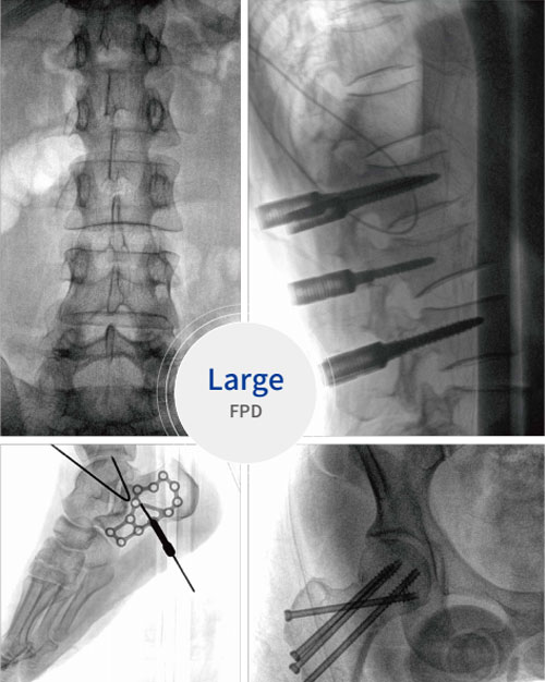
The field of view (FOV) is increased by 100% compared with the 9-inch flat panel, which helps the surgeon to quickly localize the vertebrae and the surgical site, avoiding multiple positioning and repeated exposures due to insufficient FOV.
Ultra-wide Monitor
The 32-inch monitor has a real-time/reference dual screen, making it easy to read the high-resolution film.
4-Megapixel Imaging
Equipped with 4-megapixel flat panel detector, the 3D C-arm makes clinical images clearer and easy to observe tiny lesions, ensuring high-quality imaging of complex bonelacuna and soft tissues, such as chest, abdomen, spinal joints and so on.
Intelligent Image Post-processing
The workstation system processes the captured images and can intelligently recognize human tissues and metal implants. In addition, the system processes the image through artifact processing, tissue equalization, recursive noise reduction, etc., so as to present the images of surgical regions of interest (ROI) clearly.
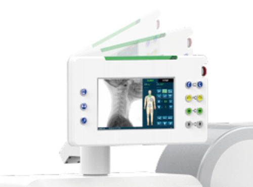
Touch Screen
Some functions can be conveniently operated, such as exposure parameters adjustment,motorized movement, and image post-processing. The touch screen supports large angle rotation, easy to operate.
5D Motorized Movements
The C-arm supports multi-dimensional movements to meet complex positioning requirements, and motorized control makes the operation convenient and safe.

Concealed cables
All cables around the C-arm are built-in, making wiping and cleaning easier. The unit can be always kept clean, and it reduces cleaning time after surgery.
Adjustable SID
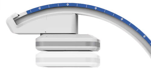
The FPD can be flexibly adjusted, raised to increase the opening distance of the C-arm and lowered to get closer to the exposed area to expand the FOV.
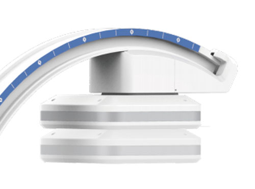
Large Free Space
The maximum space of 94 cm meets the positioning needs of obese patients or thicker tissues and reserves sufficient operation space for the surgeon.
Removable Grid
For dose-sensitive people (pediatric, etc), the filter grid can be easily and manually pulled out, which can save time and reduce the exposure dose.


It is able to adjust and preview the FOV in a ray-free state to reduce radiation damage from unnecessary exposure testings.

The intelligent DAP monitoring system can record the single examination dose and manage the radiation absorption of patients.

Low dose mode can significantly reduce the dose intake and unnecessary radiation, suitable for a variety of clinical examination needs.

Four-mode additional metal filters are available to effectively filter soft X-rays from the initial radiation, improving imaging clarity while reducing patient radiation absorption.

The model has automatic adjustment of dose parameters according to the body parts. It can reduce radiation absorption while ensuring high-quality images.

Leakage radiation in the loading state of X-ray tube is far more lower than CFDA and FDA standard, providing better protection from the X-ray source.

Pulsed X-rays are radiated at high power to obtain high-contrast images at the minimized radiation dose.
Integrated with a standardized data transmission interface for navigation or robotic systems,
PLX C7600 series is compatible with the mainstream navigation or robotic systems on the market.
Navigation or robotic systems require the input of intraoperative 3D images, which can be seamlessly transmitted to the navigation system after image acquisition, containing high-resolution
3D anatomical data as well as spatial coordinate information, so that the navigation system can perform subsequent operations with image guidance.
The combination of 3D images and navigation and positioning systems can accurately complete implant positioning, especially in complex minimally invasive surgery, which can effectively reduce surgical incisions and bleeding, reduce the need for revision surgery and postoperative CT scans, and improve surgical efficiency.
