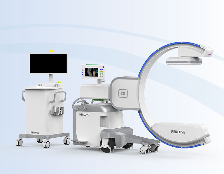
Wireless Mobile FPD C-arm Wireless Mobile FPD C-arm
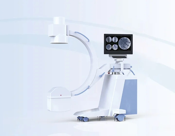
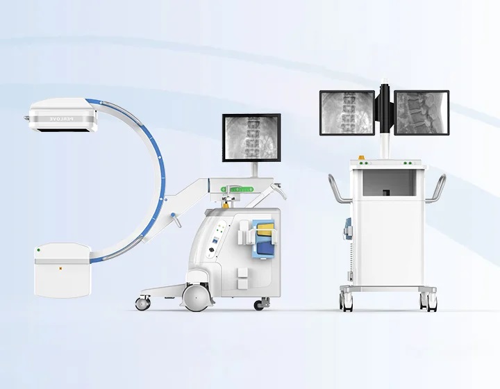
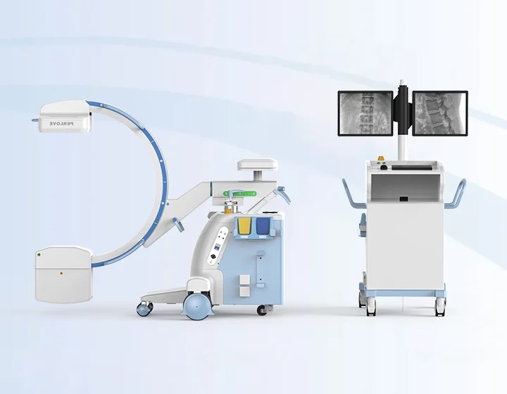
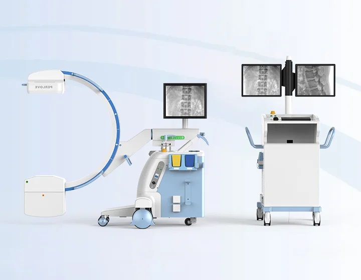
Wireless Freedom, Seamless EfficiencyWireless Connection, OR-Friendly No cables are required between the C-arm and the workstation. Information is transmitted via wireless signals, enabling faster and more flexible movement of the equipment, while eliminating safety hazards associated with cable clutter and tripping. Fast and Stable Data Transmission The system is equipped with a high-performance wireless transmission module, featuring strong anti-interference capability, wide coverage, high transmission speed, and stable signal transmission.
| Convenient, Flexible, EfficientConvenient Operation, Fast Positioning Both the tube and flat panel feature a laser positioning system for efficient intraoperative positioning without radiation exposure. It reduces radiation dose for both patients and medical staff while enhancing clinical work efficiency. Intelligent Image Processing Equipped with intelligent real-time image processing, the system automatically analyzes, enhances and optimizes images. It eliminates tedious manual operations and quickly delivers high-quality images.
|
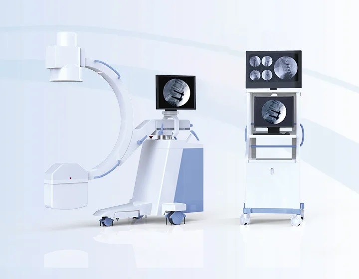
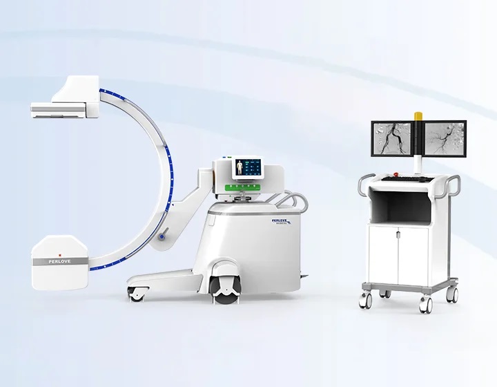
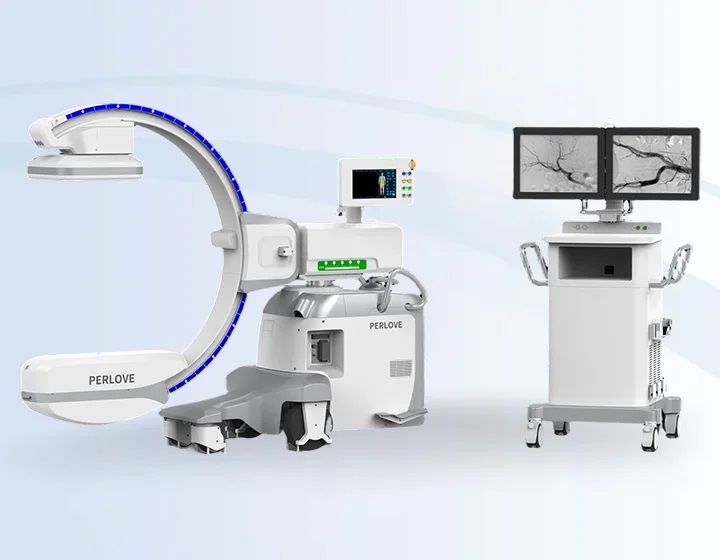
Upgraded FPDExpanded Field of View: Higher Efficiency, Lower Radiation An optional large-size dynamic flat panel detector (FPD) doubles the imaging area. It meets the needs of large-field-of-view surgeries, helping clinicians accurately locate lesions and optimize surgical planning. The elimination of repeated fluoroscopy caused by limited field of view not only improves surgical efficiency, but also effectively reduces radiation dose. Innovative Material: Advanced Imaging The optional IGZO flat panel features higher electron mobility for smooth, blur-free imaging. With finer pixel design, image resolution is significantly enhanced to clearly visualize anatomical details. Meanwhile, it greatly improves Detective Quantum Efficiency (DQE).
|
|
Precision in Details
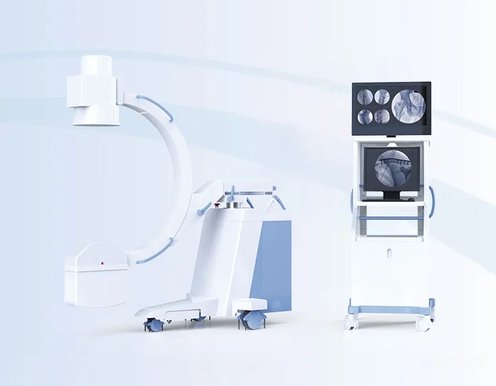
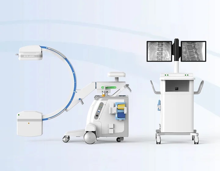
Articulating Monitors Large-angle rotatable monitors support multi-directional adjustment, delivering versatile observation perspectives for precise clinical operations.
| Mobile Workstation Cart The easily maneuverable workstation canbe placed outside the operating room to enable remote exposure, effectively minimizing radiation.
X-ray Safety Lock When the lock is engaged, no X-ray emission occurs, eliminating the risk of accidentalexposure from unintended foot switch activation. Handheld Controller The handheld remote controller enables C-arm movement and exposure parameter adjustment, streamlining clinical procedures for enhanced efficiency. |
Dynamic Flat Panel Detector The dynamic FPD delivers high DQE, low noise, wide dynamic range, excellent imaging performance and clear images. Removable Grid For pediatric and other vulnerable patient groups, the grid can be easily removed to minimize radiation dose.
Monoblock X-ray Tube High-quality monoblock X-ray generator. | Multiple Protection Reduced RadiationLower Leakage Radiation in the Loading State The X-ray source features a highly sealed design that effectively prevents X-ray leakage. When the device is in operation, radiation leakage is as low as 0.06 mGy/h, significantly enhancing radiation safety for both patients and medical staff.
DAP Real-time exposure dose is displayed on the image, enabling physicians to monitor radiation conditions and effectively control radiation dose. Proprietary High-Frequency Inverter Adopting proprietary high-frequency high-voltage generator, it features a high main inversion frequency that minimizes soft X-ray production and effectively enhances X-ray quality. Intelligent Pulsed Fluoroscopy Equipped with leading intelligent variable-frequency low-dosed pulsed fluoroscopy technology, it significantly improves imaging quality while effectively reducing radiation dose. |
Virtual Collimator The virtual collimator enables clinicians to preview and adjust the exposure field without radiation, improving workflow efficiency while minimizing unnecessary dose. Filter Design The specialized filter design effectively filters out soft rays, reducing radiation dose for enhanced safety. | Multidisciplinary Use Versatile SolutionsUnicondylar knee arthroplasty (UKA) was performed.
|
Anterior cervical decompression, bone grafting and internal fixation were performed.
Fracture reduction and pedicle screw internal fixation were performed. |
Open reduction and internal fixation with a plate and screws was performed.
|
Total hip arthroplasty was performed. Intraoperative imaging allows clear visualization of the hip joint, helping surgeons evaluate prosthesis positioning and ensure a successful procedure.
| Internal iliac artery embolization was performed. Injection of contrast agent allows clear visualization ofblood vessels, helping surgeons perform precise embolization of the internal iliac artery, control bleeding and save the patient’s life.
|
Wireless Connection, OR-Friendly No cables are required between the C-arm and the workstation. Information is transmitted via wireless signals, enabling faster and more flexible movement of the equipment, while eliminating safety hazards associated with cable clutter and tripping. Fast and Stable Data Transmission The system is equipped with a high-performance wireless transmission module, featuring strong anti-interference capability, wide coverage, high transmission speed, and stable signal transmission. Convenient Operation, Fast Positioning Both the tube and flat panel feature a laser positioning system for efficient intraoperative positioning without radiation exposure. It reduces radiation dose for both patients and medical staff while enhancing clinical work efficiency. Intelligent Image Processing Equipped with intelligent real-time image processing, the system automatically analyzes, enhances and optimizes images. It eliminates tedious manual operations and quickly delivers high-quality images. Expanded Field of View: Higher Efficiency, Lower Radiation An optional large-size dynamic flat panel detector (FPD) doubles the imaging area. It meets the needs of large-field-of-view surgeries, helping clinicians accurately locate lesions and optimize surgical planning. The elimination of repeated fluoroscopy caused by limited field of view not only improves surgical efficiency, but also effectively reduces radiation dose. Innovative Material: Advanced Imaging The optional IGZO flat panel features higher electron mobility for smooth, blur-free imaging. With finer pixel design, image resolution is significantly enhanced to clearly visualize anatomical details. Meanwhile, it greatly improves Detective Quantum Efficiency (DQE). Articulating Monitors Large-angle rotatable monitors support multi-directional adjustment, delivering versatile observation perspectives for precise clinical operations. Mobile Workstation Cart The easily maneuverable workstation canbe placed outside the operating room to enable remote exposure, effectively minimizing radiation. X-ray Safety Lock When the lock is engaged, no X-ray emission occurs, eliminating the risk of accidentalexposure from unintended foot switch activation. Handheld Controller The handheld remote controller enables C-arm movement and exposure parameter adjustment, streamlining clinical procedures for enhanced efficiency. Dynamic Flat Panel Detector The dynamic FPD delivers high DQE, low noise, wide dynamic range, excellent imaging performance and clear images. Removable Grid For pediatric and other vulnerable patient groups, the grid can be easily removed to minimize radiation dose. Monoblock X-ray Tube High-quality monoblock X-ray generator. Lower Leakage Radiation in the Loading State The X-ray source features a highly sealed design that effectively prevents X-ray leakage. When the device is in operation, radiation leakage is as low as 0.06 mGy/h, significantly enhancing radiation safety for both patients and medical staff. DAP Real-time exposure dose is displayed on the image, enabling physicians to monitor radiation conditions and effectively control radiation dose. Proprietary High-Frequency Inverter Adopting proprietary high-frequency high-voltage generator, it features a high main inversion frequency that minimizes soft X-ray production and effectively enhances X-ray quality. Intelligent Pulsed Fluoroscopy Equipped with leading intelligent variable-frequency low-dosed pulsed fluoroscopy technology, it significantly improves imaging quality while effectively reducing radiation dose. Virtual Collimator The virtual collimator enables clinicians to preview and adjust the exposure field without radiation, improving workflow efficiency while minimizing unnecessary dose. Filter Design The specialized filter design effectively filters out soft rays, reducing radiation dose for enhanced safety. Unicondylar knee arthroplasty (UKA) was performed. Anterior cervical decompression, bone grafting and internal fixation were performed. Fracture reduction and pedicle screw internal fixation were performed. Open reduction and internal fixation with a plate and screws was performed. Total hip arthroplasty was performed. Intraoperative imaging allows clear visualization of the hip joint, helping surgeons evaluate prosthesis positioning and ensure a successful procedure. Internal iliac artery embolization was performed. Injection of contrast agent allows clear visualization ofblood vessels, helping surgeons perform precise embolization of the internal iliac artery, control bleeding and save the patient’s life.Wireless Freedom, Seamless Efficiency

Convenient, Flexible, Efficient

Upgraded FPD


Precision in Details


Multiple Protection Reduced Radiation


Multidisciplinary Use Versatile Solutions
Clinical imaging helps physicians evaluate knee joint lesions and guide accurate prosthesis placement. Postoperatively, the prosthesis is stable with no complications.
Clinical images allow clear visualization of the cervical spine contour and detailed lesions, assisting in surgical planning and reducing intraoperative complications.
Guided by intraoperative imaging, surgeons can promptly adjust the approach angle and accurately place screws into thevertebral body through the pedicle, greatly improving surgical success rates.
Intraoperative imaging assists surgeons in achieving anatomical reduction, helping avoid postoperative complications caused by inadequate reduction.