

PLX 119C-CE Certification

WIDER FIELD OF VIEW |  |
Simplicity revealed in every details |
Upgraded FPD |
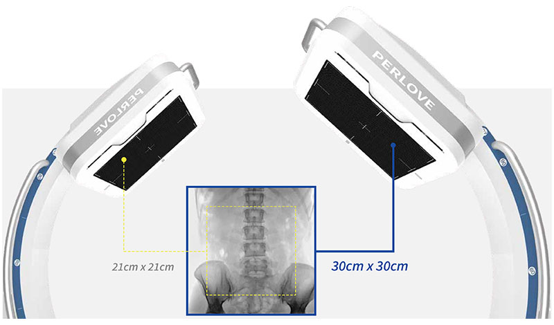
Larger field of view Equipped with a 30cm*30cm large-size dynamic FPD, imaging area has been increased by 100% compared with the conventional FPD(Optional 21cm FPD).
Crystal-clear image with delicacy 4MP resolution image combined with low signal-noise ratio reveal the tiniest anatomical details. |
Maximize Effective AreaMinimize Exposure Time
5 lumbar vertebrae can be photographed within one image by a 30cm x 30cm FPD, largely decreasing exposure times caused by insufficient image FOV, making it easier for surgeons to locate lesions and to make surgical plans in the procedure like pedicle screw fixation or procedure with strict imaging demand. |
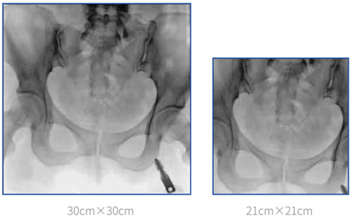
lmage taken by 30cm x 30cm FPD can basicly cover the entire pelvic floor. In the surgery of bilateral-pevic fractures or pelvic ring internal fixation, all the fracture sites can be revealed by single exposure, which significantly improves overall efficiency. |
Clinical image
Clinical application
Joint surgery lmage with high resolution can clearly reveal articular structure, helping surgeons to better locate the target. |
Trauma surgery Flexible mechanical movement realize easy positioning for different type of trauma surgery, providing comfortable handling experience.
Spine surgery upgraded FPD present more details of vertebrae, helping surgeons to better making surgical plans.
Pain management Multiple radiation protection decrease overall exposure times during minimally invasive procedure.
|
Low dose management |
High frequency pulsed fluoroscopy X-ray generated in high frequency pulse wave can decrease 50% of radiation dose compared with a continous fluoroscopy.
DAP monitoring Real-time DAP displayed on the screen.
Intelligent parameter adjustment Optimized and adjustable exposure parameter created by intelligent hardware and algorithm provides a friendly examination scenario.
Virtual collimator Allowing radiologists to preview and to adjust exposure area before exposure, reducing unnecessary radiation.
Aluminum beam filtration Decreasing radiation level by absorbing soft X-ray. Ensuring high quality image for precise diagnosis and treatment. |
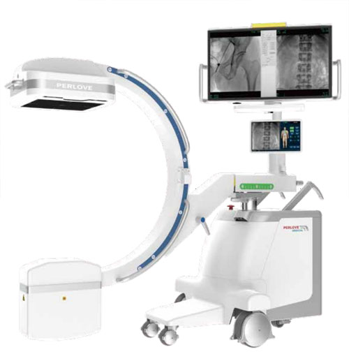
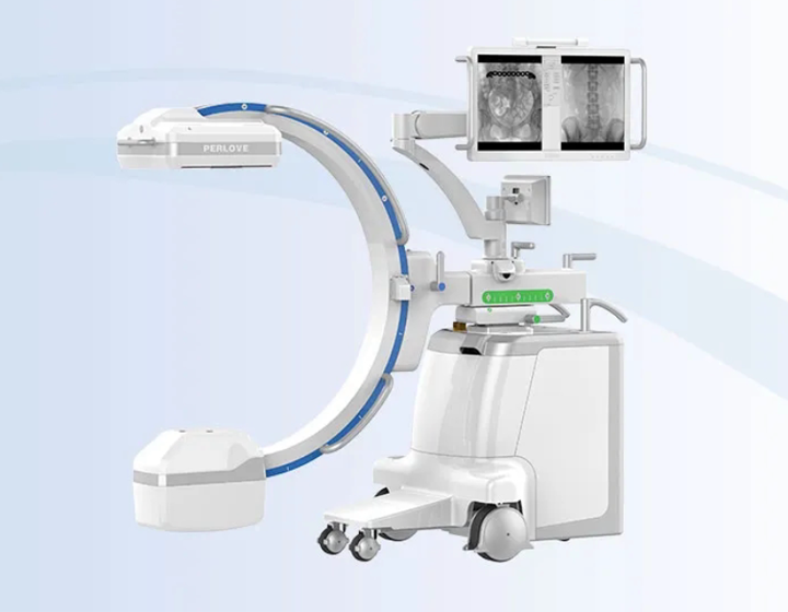
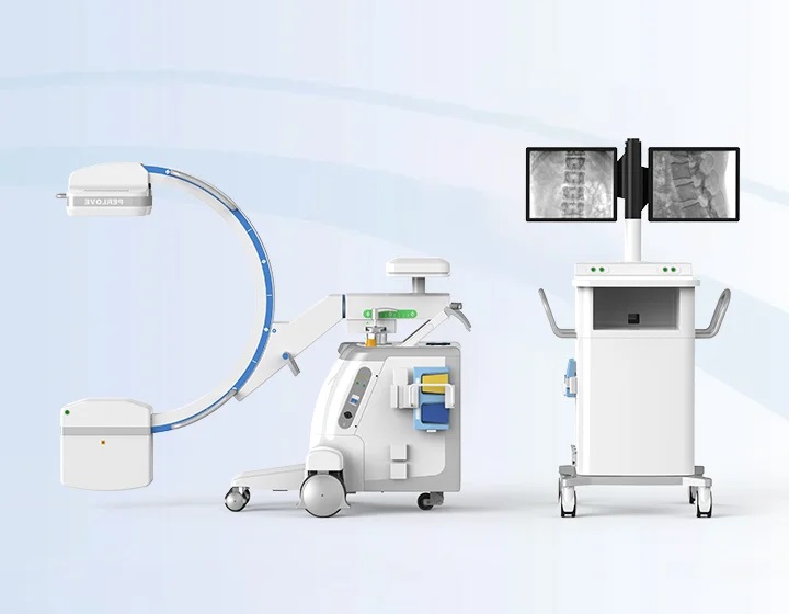
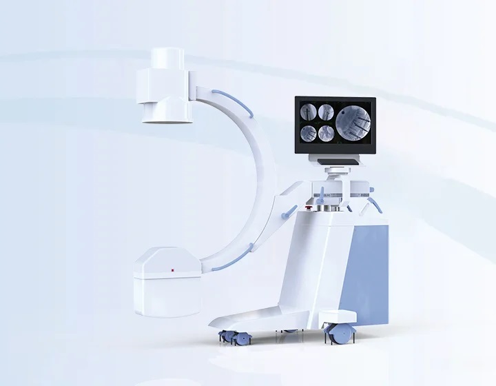
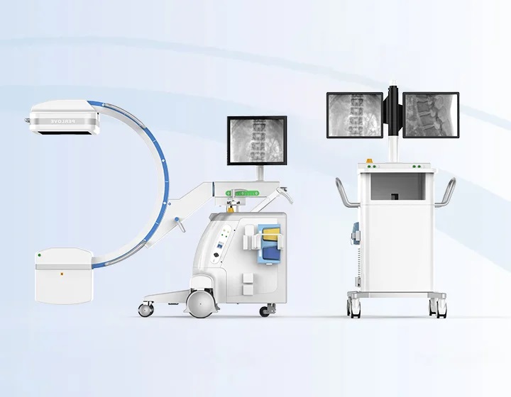
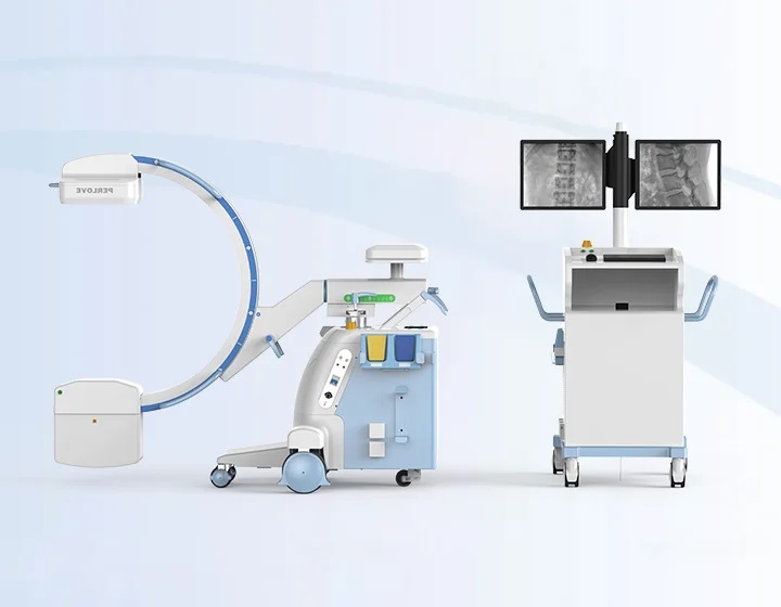
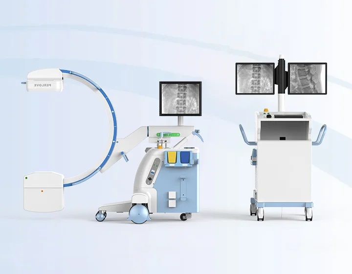
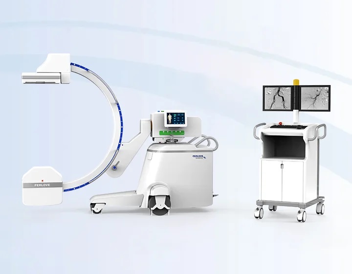
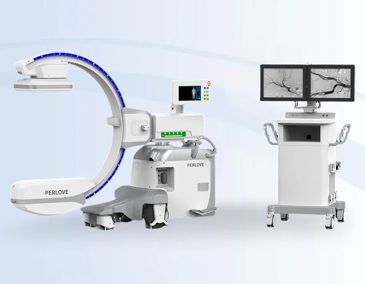
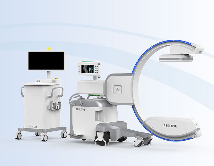
All-in-one Compact Design Flexible and EfficientCompact design + Standby power = A seamless connection among ORs. More convenient operation · The integrated workstation integrates the image acquisition system and the X-ray generator control system, which is convenient and quick to operate. Easy and flexible movement · The allin-one compact design of C-arm eliminates the monitor cart and wires, which makes it move flexibly. The machine can be easily carried out by operators with one hand. Recovery management ·The time to get all the functions of the equipment operable should not exceed 5 minutes.When AC power is accidentally lost, the system will be unusable. The workstation computer and LCD are powered by UPS, and a message is displayed on the LCD informing the operator that the system will shut down within 30 seconds. ·After the system is restored to power, press the power on button to start the system and the computer will automatically tum on. Wait for the workstation software to start automatically, a user dialog box will pop up, select the username, enter the password, and start using. | User-friendly Touch Interface
Easy exposure control Equipped with the high-quality and large-size touchscreen, high sensitivity, flexible rotation, more convenient for clinicians to perform touch operation. Large-size monitor Dual screen interface is convenient for surgeons to obtain more comprehensive clinical information Clear clinical images The sharp clinical images can be guaranteed. It can realize clear display of tiny anatomical structures, with no loss of image information, bringing more comfortable reading for surgeons. |
Application Intended for use by a qualified/trained doctor or technologist on both adult and pediatric patients for taking diagnostic radiographic exposures of the skull, spinal column, chest, abdomen, extremities, and other body parts.Applications can be performed with patient sitting, standing or lying in the prone or supine positions. Not for mammography. |
Technical Specifications
Nominal X-ray tube voltage | 125kVp |
Nominal focus size | 0.6/1.8 |
Maximum anode heat content | 35.5kJ |
The angle of circular sliding along the track of C-arm | 130°~140°
|
The vertical movement range of column | 380 mm~420 mm |
The horizontal movement range of C-arm | 190 mm~210 mm |
Safety information Pregnant women and breast-feeding women are not suitable.Be sure to take precautions from X-ray before each exposure for patient and user. As permitted by radiography, the exposure shall be conducted with as low dose as possible.As radioactive equipment, this equipment shall be operated by operator strictly according to provisions of the User Manual and cannot be used for other purpose except medical radiography. |


Larger field of view
Equipped with a 30cm*30cm large-size dynamic FPD, imaging area has been increased by 100% compared with the conventional FPD(Optional 21cm FPD).

Crystal-clear image with delicacy
4MP resolution image combined with low signal-noise ratio reveal the tiniest anatomical details.

Full lumbar vertebrae imaging
5 lumbar vertebrae can be photographed within one image by a 30cm x 30cm FPD, largely decreasing exposure times caused by insufficient image FOV, making it easier for surgeons to locate lesions and to make surgical plans in the procedure like pedicle screw fixation or procedure with strict imaging demand.

Entire pelvic imaging
lmage taken by 30cm x 30cm FPD can basicly cover the entire pelvic floor. In the surgery of bilateral-pevic fractures or pelvic ring internal fixation, all the fracture sites can be revealed by single exposure, which significantly improves overall efficiency.


Joint surgery
lmage with high resolution can clearly reveal articular structure, helping surgeons to better locate the target.

Trauma surgery
Flexible mechanical movement realize easy positioning for different type of trauma surgery, providing comfortable handling experience.

Spine surgery
upgraded FPD present more details of vertebrae, helping surgeons to better making surgical plans.

Pain management
Multiple radiation protection decrease overall exposure times during minimally invasive procedure.


Lower leakage radiation in the loading state
Leakage radiation in the loading state of X-ray tube is far more lower than CFDA and FDA standard, providing better protection from the X-ray source.


High frequency pulsed fluoroscopy
X-ray generated in high frequency pulse wave can decrease 50% of radiation dose compared with a continous fluoroscopy. This technology has a positive effect on weaking the motion artifact.

DAP monitoring
Real-time DAP displayed on the screen.
Easy for monitoring radiation level.

Intelligent parameter adjustment
Optimized and adjustable exposure parameter created by intelligent hardware and algorithm provides a friendly examination scenario.

Virtual collimator
Allowing radiologists to preview and to adjust exposure area before exposure, reducing unnecessary radiation.

Aluminum beam filtration
Decreasing radiation level by absorbing soft X-ray. Ensuring high quality image for precise diagnosis and treatment.
Compact design + Standby power = A seamless connection among ORs.
More convenient operation
· The integrated workstation integrates the image acquisition system and the X-ray generator control system, which is convenient and quick to operate.
· When surgeons perform complex surgeries,it is easy for the surgeon to observe the surgical results and monitor the surgical process in time.
Easy and flexible movement
· The allin-one compact design of C-arm eliminates the monitor cart and wires, which makes it move flexibly. The machine can be easily carried out by operators with one hand.
Recovery management
·The time to get all the functions of the equipment operable should not exceed 5 minutes.When AC power is accidentally lost, the system will be unusable. The workstation computer and LCD are powered by UPS, and a message is displayed on the LCD informing the operator that the system will shut down within 30 seconds.
·After the system is restored to power, press the power on button to start the system and the computer will automatically tum on. Wait for the workstation software to start automatically, a user dialog box will pop up, select the username, enter the password, and start using.

Easy exposure control
Equipped with the high-quality and large-size touchscreen, high sensitivity, flexible rotation, more convenient for clinicians to perform touch operation.
Large-size monitor
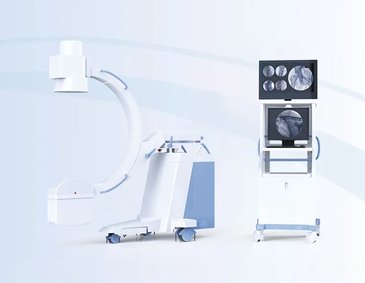
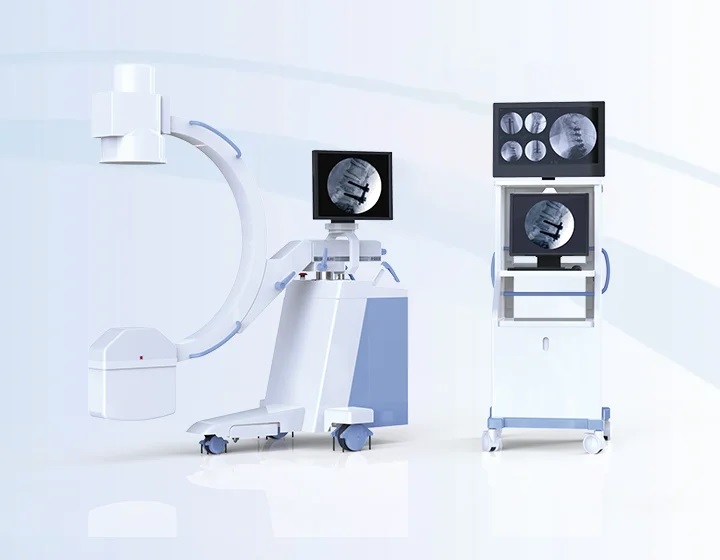
Dual screen interface is convenient for surgeons to obtain more comprehensive clinical information
Clear clinical images
The sharp clinical images can be guaranteed. It can realize clear display of tiny anatomical structures, with no loss of image information, bringing more comfortable reading for surgeons.
Application
Intended for use by a qualified/trained doctor or technologist on both adult and pediatric patients for taking diagnostic radiographic exposures of the skull, spinal column, chest, abdomen, extremities, and other body parts.
Applications can be performed with patient sitting, standing or lying in the prone or supine positions. Not for mammography.
Technical Specifications
Nominal X-ray tube voltage | 125kVp |
Nominal focus size | 0.6/1.8 |
Maximum anode heat content | 35.5kJ |
The angle of circular sliding along the track of C-arm | 130°~140°
|
The vertical movement range of column | 380 mm~420 mm |
The horizontal movement range of C-arm | 190 mm~210 mm |
Safety information
Pregnant women and breast-feeding women are not suitable.Be sure to take precautions from X-ray before each exposure for patient and user. As permitted by radiography, the exposure shall be conducted with as low dose as possible.As radioactive equipment, this equipment shall be operated by operator strictly according to provisions of the User Manual and cannot be used for other purpose except medical radiography. As improper operation will damage this equipment, it can only be used by trained personnel with needed radiation knowledge.